Article Text

Abstract
Multiple primary tumors can be classified as synchronous or metachronous. Cases have been reported, with a prevalence, in gynecologic malignancies, of 1.9 to 4.3%, and commonly occurring in endometrial and ovarian malignancies. Renal tumors coexisting with primary cervical cancer are mostly metastatic tumors, and at present, no case of cervical carcinoma metachronous with renal cell carcinoma has been reported on literature.
This is a case of Papillary Squamous Cell Carcinoma of the cervix who developed a metachronous Clear Cell Renal Cell Carcinoma. Several months after the diagnosis of cervical cancer, she presented with an abdominal mass and signs of uremia secondary to obstructive uropathy. She underwent radical nephrectomy with contralateral percutaneous nephrostomy. Definitive plan for the cervical mass is concurrent chemotherapy and radiation, depending on the improvement in renal function.
Currently, there are no clearly established guidelines in managing metachronous cervical and renal masses, and this presents a unique opportunity to document this case, and study its implications on management and prognosis.

A. Low Power Objective (100x magnification) of Papillary Squamous Cell Carcinoma with tumor cells arranged in nests and papillary configuration. B. High Power Objective (400x). These tumor cells exhibit moderate pleomorphism, with enlarged, round to oval, nuclei, some with prominent nucleoli, and abundant eosinophilic cytoplasm and distinct cell borders

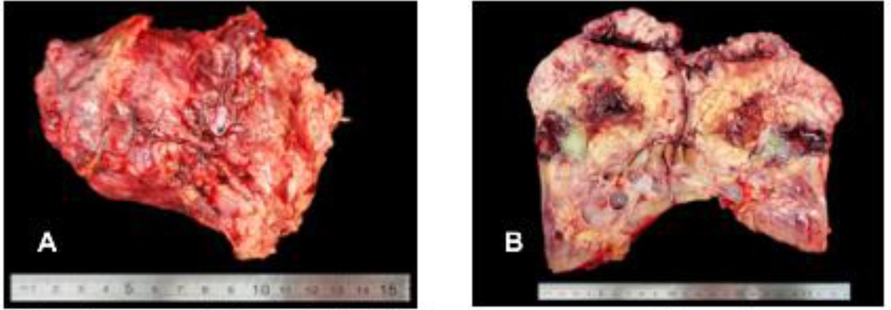
Intraoperative pictures. A. The left kidney was converted to a solid, necrotic mass measuring 12.0 × 10.0 × 5.0 cm. B. Cut section of the resected left kidney

A. Low Power Objective (100x magnification) of Clear Cell Renal Cell Carcinoma, ISUP Grade 1, with tumor cells distributed in alveolar pattern, separated by thin blood vessels. B. High Power Objective (400x). These tumor cells exhibit moderate pleomorphism, with hyperchromatic nuclei, rare nucleoli, and clear cytoplasm