Article Text

Abstract
Aims The International Endocervical Adenocarcinoma Criteria and Classification (IECC) was recently proposed as an improved method for categorising endocervical adenocarcinoma (EA) into human papillomavirus (HPV)–associated adenocarcinomas (HPVAs) and non-HPV-associated adenocarcinomas (NHPVAs). Such categorisation correlates with patient age and tumour size; however, its association with patient outcome remains to be established.
Methods Institutional cases of EA with histological material available were selected. Three gynaecological pathologists independently classified all tumours according to the IECC with consensus review used when necessary. Clinicopathologic variables were recorded for each case.
Results Of a total of 87 EAs, 71 (82%) were classified as HPVA and 16 (18%) as NHPVA. Among HPVA, most were usual type (51/71, 72%) followed by mucinous not otherwise specified (10/71, 14%) and invasive stratified mucin-producing carcinoma (ISMC, 8/71, 11%). Most NHPVAs were of gastric type (12/16, 71%) followed by clear cell and mesonephric (two each, 12%). Compared with HPVAs, NHPVAs were significantly associated with older age (p<0.001), larger horizontal extent (p=0.013), greater depth of invasion (p=0.003), lymphovascular space invasion (p<0.001), advanced stage (p<0.001) and invasive pattern C (p<0.001). On univariate analysis, worse disease-free survival (DFS) and disease-specific survival (DSS) correlated with NHPVA group. Among the HPVA subtypes, ISMC showed worse DFS and DSS compared with other HPVA types.
Conclusions The simple morphological approach of the IECC appears to be prognostically valuable. NHPVA (in particular gastric type) and ISMC (a recently recognised subset of HPVA) have an adverse outcome and their recognition following the IECC is important. We provide further evidence to replace the current WHO classification with the IECC.
- Gynecological pathology
- cervical cancer
- HPV
- cervix
Statistics from Altmetric.com
Introduction
Endocervical adenocarcinoma (EA), the second most common malignancy of the uterine cervix, has been noted to have an increasing incidence worldwide.1 2 A large proportion of EAs are associated with oncogenic human papillomavirus (HPV) infection; however, it is well recognised that a proportion of EAs are not related to HPV infection.3–9 Studies which have evaluated the clinical behaviour and patient outcomes of gastric-type EA (the most common type of non-HPV-related adenocarcinoma, NHPVA) have shown poor prognosis compared with usual-type EA (the most common type of HPV-related adenocarcinoma, HPVA).10–12
According to the 2014 World Health Organization (WHO) classification of tumours of female reproductive organs,13 EAs are subclassified based mostly on descriptive characteristics focused on cytoplasmic features. Limitations of this classification system have been recognised, as the definitions provided are subjective and not linked to clinical behaviour or aetiology. In addition, such morphological stratification is largely ignored by clinicians when planning patient management.14 Recently, an international group of gynaecological pathologists endeavoured to establish the International Endocervical Adenocarcinoma Criteria and Classification (IECC), a scheme supported by aetiology and based on the presence or absence of HPV infection–related morphological features.15 Aetiology-based classification schemes have been successfully implemented and used in other organs such as the vulva16–19 and oropharynx.20 21 The seminal study by Stolnicu et al introducing the IECC showed that (1) EAs can be reliably segregated into HPVA and NHPVA based on the presence or absence of easily identifiable apical mitoses and apoptotic bodies (HPV infection–related features), (2) HPVA and NHPVA groups can both be further subclassified based on existing morphologicl criteria, and (3) the IECC is supported by immunohistochemistry and HPV in situ hybridisation (ISH) results.15 By virtue of its simplified and practical approach, we recently demonstrated that the IECC has improved interobserver agreement compared with the WHO classification.22 Moreover, we also documented excellent correlation between IECC grouping and HPV status.22
Under the IECC, HPVA and NHPVA differ significantly in terms of patient age and tumour size,15 but correlation with other important clinical and pathological variables including patient outcome remains to be established. We hypothesised that IECC grouping correlates with significant clinical and pathological risk factors and with patient survival. Thus, we sought to comprehensively evaluate the clinicopathological features of a cohort of patients with EA in the context of the novel IECC classification.
Methods
Case selection and review
Cases with a final diagnosis of invasive EA and adenosquamous carcinoma over a 16-year period (January 2002–December 2017) were identified through a systematic search of the Sunnybrook Health Sciences Centre Pathology Information System. The search included resection (cone biopsy, loop electrosurgical excision procedure, trachelectomy or hysterectomy) and biopsy specimens. Those with histological material available for examination were further reviewed, and complete slide sets for each case were examined by one gynaecological pathologist (CP-H) in order to confirm the diagnosis of EA. Tumours initially diagnosed as adenosquamous carcinoma but reclassified on review by the senior author as EA were also included, and confirmed cases of adenosquamous carcinoma were excluded. Tumours of uncertain origin and metastases to the cervix were also excluded. The following data were recorded for each case: type of specimen (biopsy or resection), date of diagnosis, age at diagnosis and, if applicable, horizontal extent, depth of invasion, margin status, pattern of invasion as defined by the Silva system,23 presence or absence of lymphovascular invasion and lymph node metastases, International Federation of Gynecology and Obstetrics (FIGO) stage, neoadjuvant or adjuvant treatment status, date of last known follow-up, date of recurrence, date of death and cause of death.
IECC categorisation
Selected histopathological material (two to four slides) from each case was reviewed by three gynaecological pathologists (CP-H, BEH and MRN) who independently assigned a diagnosis according to IECC morphological criteria.15 Of note, these pathologists are experienced in applying in the IECC and participated in a recent interobserver and validation study evaluating its application.22 As part of the previous study, written instructions regarding classification were provided, asking the reviewer to first establish the presence or absence of easily identifiable apical mitotic figures and apoptotic bodies. When these features were seen, the tumour was designated as a HPVA. Conversely, the diagnosis of NHPVA was assigned if mitoses and apoptosis were not abundant or easily identified. HPVAs were further subcategorised based on traditional cytoplasmic and architectural features as follows: usual type (intracellular mucin in <50% of tumour cells), mucinous (intracellular mucin in >50% of cells; further subcategorised as mucinous not otherwise specified (NOS), intestinal or signet-ring cell), villoglandular or invasive stratified mucin-producing adenocarcinoma (ISMC). NHPVA were also subcategorised based on existing morphological criteria as gastric type (including minimal deviation adenocarcinoma), clear cell, mesonephric, endometrioid or serous. Cases not amenable for categorisation were deemed as EA-NOS. Examples of HPVA and NHPVAs are depicted in figures 1 and 2, respectively.
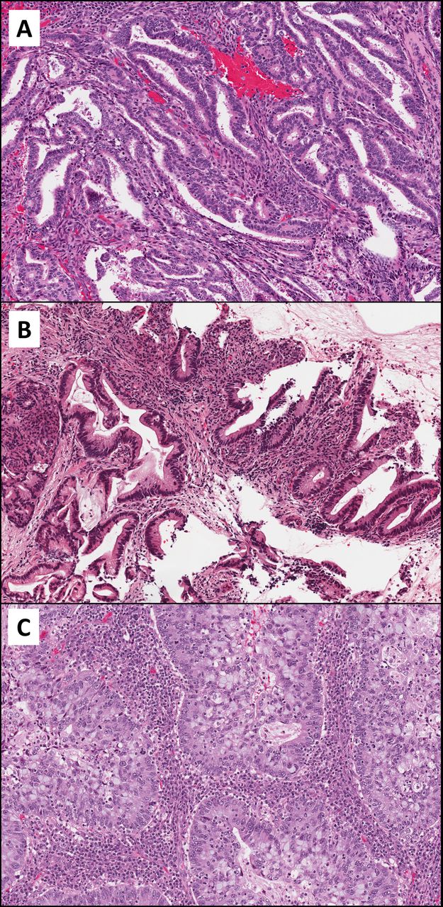
Examples of human papillomavirus–associated endocervical adenocarcinoma. (A) Usual type. (B) Mucinous, not otherwise specified. (C) Invasive stratified mucin-producing carcinoma. Note the presence of apical mitoses and apoptotic bodies in all three images.

Examples of human papillomavirus–unrelated endocervical adenocarcinoma. (A) Gastric type. (B) Clear cell type. (C) Mesonephric type.
After the independent case review step, the final diagnostic category was assigned. Fully concordant diagnoses (agreement by all three pathologists) required no further review. Discrepant cases were assigned a majority diagnosis (when two reviewers concurred). Instances of complete discrepancy among the three reviewers were not encountered. All reviewers were blinded to all clinicopathological characteristics as well as immunohistochemistry and HPV ISH and/or PCR results (if existing).
Ancillary studies
Final IECC diagnosis were compared with immunohistochemical results for p16 (clone JC8, Ventana) and p53 (clone DO-7, Ventana) which were performed in whole-tissue sections or tissue microarrays constructed using two 2 mm cores of representative tumour tissue per case. Normal tissue controls were included in each tissue microarray block. Stains were interpreted by one of the authors (CP-H) and recorded as follows: p16 staining was considered ‘overexpressed’ if diffuse block-like nuclear and cytoplasmic staining was seen in ≥80% of tumour cells, ‘patchy’ if positivity was patchy (<80% of tumour cells) or cytoplasmic only, or ‘absent’ if no staining was seen; p53 was scored as ‘overexpressed’ if ≥80% of tumour nuclei were strongly positive, ‘null’ when no staining was seen in tumour cells in the presence of an intact internal control or ‘wild type’ when nuclear expression was heterogeneous in intensity. HPV ISH was performed in cases represented in tissue microarrays; methodology and results were described in a previous study.22
Statistical analysis
For analysis purposes, time of recurrence and time of death were used to estimate disease-free survival (DFS) and disease-specific survival (DSS). The time to recurrence was defined from the date of primary surgery to the date of first recorded recurrence by imaging or tissue diagnosis. Likewise, time to death was defined from the date of primary surgery. DSS counted exclusively events of cancer-related death. Univariate DFS and DSS probabilities were estimated and plotted using Kaplan-Meier method and compared using the log-rank test. Cox proportional-hazards models were used to generate univariate HRs and 95% CIs to analyse individual contributions of each recorded clinical and pathological variable to disease-free and disease-specific probabilities. Multivariate Cox proportional-hazards regression model analysis was subsequently conducted to weigh clinically meaningful variables that were significant on univariate analysis. The correlation between tumour type and other clinicopathological characteristics was calculated using appropriate statistical tests, that is, χ2 test for categorical variables and Mann-Whitney U test for continuous variables. All statistical analyses were performed using the SPSS software V.24.0 (IBM). Two-sided p values less than 0.05 were considered statistically significant.
Results
A total of 90 consecutive patients met inclusion criteria. Morphological classification according to the IECC separated the cohort into 71 HPVAs (78%), 16 NHPVAs (19%) and 3 EA-NOS (3%). The latter group was excluded from further analysis. Among the 87 EAs included in the final cohort, usual-type HPVA was the most common subtype comprising 59% (51/87) of the series. Gastric-type NHPVA (14%, 12/87) and mucinous NOS HPVA (11%, 10/87) were the second and third most common subtypes, respectively. ISMC followed in frequency (8/87, 9%). Clear cell NHPVA (2/87, 2%), intestinal-type mucinous HPVA (2/87, 2%) and mesonephric NHPVA (2/87, 2%) comprised the remaining minority of cases. None of the reviewed cases were assigned to the villoglandular HPVA, signet-ring cell HPVA, serous NHPVA and endometrioid NHPVA categories (table 1). Six cases had an archival diagnosis of adenosquamous carcinoma, but on review were reclassified as EA by the initial reviewer and included in the series. All six tumours were diagnosed as HPVA by the additional pathologists: three were classified as usual type, two as ISMC and one as mucinous NOS.
Distribution of patients with endocervical adenocarcinoma classified according to the IECC
Correlation of IECC final diagnosis with immunohistochemical and HPV molecular data was high. Among the 71 HPVA tumours, 64/67 (96%) with p16 available showed overexpression of this marker; the remaining 3/67 (4%) were patchy or negative. One of these three cases was positive for HPV, another was negative and in the third case HPV testing was not performed. Likewise, 64/65 (98%) HPVAs with HPV ISH or PCR results were positive and 1/65 (2%) was negative. Conversely, among the 16 NHPVA tumours in the group, 16/16 (100%) were negative or patchy for p16, and 9/9 (100%) tested for HPV ISH were negative. 66/66 (100%) HPVAs tested for p53 were normal (wild type); in contrast, 4/14 (29%) NHPVAs with p53 available showed an abnormal staining pattern (overexpression), all of which were gastric-type adenocarcinomas (mesonephric and clear cell carcinomas had normal p53 expression).
Clinicopathological characterisation
The clinicopathological features of patients with HPVA and NHPVA are compared in table 2. Compared with HPVA, NHPVA tumours were significantly associated with the following features: older age at diagnosis (p<0.001), larger horizontal extent (p=0.013), greater depth of invasion (p=0.003), presence of lymphovascular invasion (p<0.001), advanced stage (FIGO stage 2 or higher, p<0.001) and invasive pattern C (p<0.001). Differences in lymph node involvement (p=0.097) and primary treatment modality (surgery vs chemoradiation, p=0.142) were not statistically significant.
Clinicopathological features of HPVA vs NHPVA
Survival analysis
Seventy-eight cases had follow-up data available (87%, 62/71 HPVA and 16/16 NHPVA). Mean follow-up interval was 45.2 months (median 38 months, range 1–155 months). Clinical outcomes of patients with HPVA and NHPVA are depicted in table 3. Tumour recurrence occurred more frequently in NHPVA (8/16 patients, 50%) than in HPVA (9/62 patients, 15%). Likewise, cancer-related mortality was significantly higher in NHPVA (7/16 patients, 44%) than in HPVA (2/62 patients, 3%). Of note, all deaths in the series were due to carcinoma; deaths due to other causes were not identified.
Recurrence and cancer-related mortality in patients with endocervical adenocarcinoma
Within the HPVA group, adverse events most commonly occurred in patients with ISMC: four out of eight (50%) patients with ISMC recurred (two with local recurrence and two with distant recurrence), and two (25%) died of disease. No patients with usual-type EA died of their disease, and only a small proportion (7%, 3 out of 43 cases) suffered recurrence (all distant recurrence). One of the two intestinal-type mucinous EAs recurred to distant sites while one of the nine mucinous NOS EAs recurred locally.
Among patients with NHPVA, those with gastric-type EA predominated and had the largest number of adverse events (6/12 patients developed recurrence, 5/12 died of disease). Tumour recurrence in this group included locoregional recurrence in one patient, distant (lung, peritoneum, bowel) recurrence in four patients, and both local and distant tumour recurrence in two patients. Of the two patients with clear cell EA, one developed recurrence 4 months after diagnosis and had a protracted course ending in demise 81 months after initial diagnosis; the second patient was alive with no signs of recurrence after 18 months of follow-up. Of the two patients with mesonephric EA, one developed recurrence in the retroperitoneum 21 months after initial surgery and died 2 months later; the second patient was alive and well 16 months after diagnosis.
By univariate survival analysis, tumour grouping as per the IECC (ie, HPVA vs NHPVA) was the only variable predictive of DSS, significantly worse in NHPVA cases (p=0.001, HR 39.8, 95% CI 4.9 to 324.9). Likewise, the IECC correlated with DFS, with NHPVA having significantly worse DFS compared with HPVA (p<0.001, HR 6.8, 95% CI 2.6 to 17.8) (figure 3). Among HPVA subtypes, ISMC had significantly worse DFS and DSS when compared with other HPVA types (p=0.008 and 0.016, respectively). Other variables associated with worse DFS included horizontal extent (p=0.037, HR 1.04, 95% CI 1.00 to 1.08), depth of invasion (p<0.001, HR 1.16, 95% CI 1.08 to 1.25), lymphovascular space invasion (p<0.001, HR 17.4, 95% CI 3.6 to 83.4) and stage (overall p<0.001). On multivariate survival analysis, none of these features retained statistical association with DFS.

Kaplan-Meier curves stratifying patients with human papillomavirus (HPV)–related (HPV-associated adenocarcinoma, HPVA) and HPV-unrelated (non-HPV-associated adenocarcinoma, NHPVA) endocervical adenocarcinoma in terms of (A) disease-free survival and (B) disease-specific survival.
Discussion
The IECC is a novel classification scheme which divides EAs into clinically relevant categories based on easily recognisable morphological features. This effort echoes recent shifts in tumour classification in other organs based on aetiology (HPV infection vs not). Importantly, the diagnostic reproducibility of the IECC among gynaecological pathologists is acceptable and improved compared with the current WHO classification.22 Our study is the first to validate the IECC in terms of patient outcome. Most notably, of all the variables analysed, tumour grouping per the IECC was the only variable statistically predictive of DSS; however, we acknowledge that the number of NHPVA cases was small overall. Our study also demonstrates a significant association between IECC grouping and traditional clinical and pathological prognostic variables.24–28
It is important to note that the categorisation process followed the algorithm described in the seminal IECC publication, which is based purely on morphology. This rationale underscores the importance of routine H&E assessment in the evaluation of endocervical glandular neoplasia. The identification of apoptotic bodies and mitoses first in the evaluation proved to be a valuable diagnostic tool in our study. Even if the pathologist is experienced in the recognition of different EA types using conventional characteristics, we believe that these IECC features add value to the diagnostic exercise. In reality, most pathologists in routine practice will also use ancillary testing to confirm the EA histotype. Notably, under our approach, we obtained excellent correlation with confirmatory immunohistochemistry and HPV molecular results as reported previously by our group22 and in the expanded cohort of the current study. Nonetheless, one case classified as HPVA was negative for p16 overexpression and HPV ISH. This instance was also encountered in 7% of HPVAs in the seminal publication on the IECC.15 Possible explanations for such unexpected result include infection by HPV types not covered by the ISH test, as well as methylation-induced inactivation of the p16 gene and allelic loss of p16, which would lead to a negative p16 result. In cases with features of HPVA but negative p16 and/or HPV ISH, careful review is recommended in order to exclude NHPVA types and metastases, with ultimate classification as HPVA only after such possibilities have been ruled out. Immunohistochemistry and clinical correlation are instrumental in this exercise. A more recent analysis highlighted the role of immunohistochemistry in improving the IECC classification algorithm.29
Although there are no type-specific treatments in the management of EA as of yet, the IECC classification may help guide the design and development of clinical trials and tailored management strategies for the different forms of EA. Regardless of the subtype, all NHPVAs have a considerable risk of recurrence and death, with essentially half all of cases with follow-up having poor outcome. With these data in mind, it seems prudent to suggest that, at least currently, a diagnosis of NHPVA warrants a conventional therapeutic approach which includes radical trachelectomy or hysterectomy with or without lymph node sampling in early-stage cases and chemo-radiation in late-stage cases. It is important to note, however, that resistance to traditional chemotherapeutic agents has been reported in gastric-type EA.30
Among types related to HPV infection, our analysis also revealed potentially meaningful differences. Usual-type HPVA, by far the most frequent subtype of EA, had a low rate of recurrence and no cancer-specific deaths in our cohort. The same was observed for patients with mucinous NOS HPVA. In contrast, ISMC had significantly higher percentages of recurrence and cancer-related deaths. ISMC is a recently described entity, frequently observed in association with stratified mucin-producing intraepithelial lesion (SMILE), and believed to represent its invasive counterpart.31 In its original description, distant tumour recurrence was documented in 3/8 cases of ISMC, similar to what was observed in our cohort. These observations suggest that ISMC is an important variant within the spectrum of HPVA. It is important to note that ISMC usually grows in a solid or nested fashion and therefore may be classified as a moderately to poorly differentiated EA of usual type by some pathologists; however, emerging data suggest that this is a biologically important tumour subtype. On the other hand, it is conceivable that some poorly differentiated usual-type or mucinous NOS EAs in fact have ISMC morphology. Importantly, ISMC should not be confused with adenosquamous carcinoma, which has by definition a phenotypic squamous component. Nonetheless, we acknowledge that on routine diagnosis the distinction between ISMC and adenosquamous carcinoma can, at least in principle, be difficult. Indeed, many cases historically diagnosed as adenosquamous carcinoma may actually represent EA including ISMC, as demonstrated in our study (six of our cases had an archival diagnosis of adenosquamous carcinoma) and in a recent publication documenting a revised diagnosis of adenocarcinoma in 25/59 (42%) tumours formerly categorised as adenosquamous carcinoma.32
The presence of lymph node metastases did not significantly differ between HPVA and NHPVA in our study. Of note, a trend towards higher proportion of nodal involvement in NHPVA cases (25%, 2/8 cases) compared with HPVA cases (6%, 2/34 cases) was noted. The relatively low number of cases with lymph node sampling in our cohort (46%, 42/90 cases) may explain the lack of statistical association.
We chose to include both surgically resected as well as biopsy-only specimens, as the latter represents patients who undergo primary chemoradiation and no surgery, a traditionally under-represented and understudied subgroup of EA. Primary chemoradiation is usually reserved for tumours with advanced stage and as such, additional (and more abundant) surgical tissue may never become available. While we observed a correlation between stage and IECC diagnosis, the same was not true for primary treatment modality (chemoradiation vs surgical resection) and IECC grouping. This may reflect under-recognition of advanced disease, particularly in NHPVA cases, or surgeon preference to treat these rare and aggressive tumours surgically despite advanced stage.
Although the pattern-based classification system has been predominantly studied in HPV-associated tumours,33 34 recent work has studied its applicability in the evaluation of NHPVA. We showed that all NHPVA tumours in our series had destructive patterns of invasion (15 with pattern C and one with pattern B). Moreover, the pattern of invasion was associated with DFS in univariate analysis. In concordance with this finding, a recent study showed that HPVA pattern C tumours had a trend towards lower recurrence rates and better survival than NHPVA pattern C tumours.35 Lastly, lymph node metastases were only identified in tumours with pattern C in our cohort, further corroborating the association between invasive pattern and lymph node status.33
Our study is based on patients from a single institution and therefore is limited by its relatively small cohort size. Moreover, a statistical relationship between known prognostic variables, such as stage, and patient outcome was not observed likely due to our sample size. Thus, confirmation of our observations in a larger and multi-institutional study is warranted. In addition, while the distribution of our cases generally reflects the normal expected frequencies in the population,13 we acknowledge that certain EA subtypes are under-represented in our sample. As a result, our findings are heavily driven by usual-type HPVA and gastric-type NHPVA, the most common EA types, and further investigation of rare variants such as villoglandular, clear cell and mesonephric EA in larger studies is necessary. Mesonephric carcinomas of the endocervix and other gynaecological organs are increasingly recognised,36 and further insight into their biological behaviour and prognosis may be available in the near future. In addition, the limited data on the clinical outcomes of clear cell EA suggest that, at least at early stage, it may be similar to that of non-clear cell EAs.37 38 Of note, none of our cases were classified as endometrioid or serous subtypes, consistent with the prevailing thought that these subtypes are incredibly rare and arguably non-existent in the cervix.15 When encountering lesions with endometrioid and serous morphology as currently defined by the IECC, the more likely possibilities of endometrial endometrioid adenocarcinoma involving the cervix and serous carcinoma of the endometrium or upper genital tract should always be considered.
In summary, we have shown that the practical and reproducible IECC correlates with clinical and pathological variables including patient outcome. NHPVA and specific subtypes of HPVA appear to have a more aggressive clinical course. Thus, the application of this morphologically based diagnostic algorithm has the potential to offer valuable prognostic information and eventually guide treatment. Our findings highlight the importance of routinely classifying EA following the IECC and support replacing the WHO scheme with this novel system.
Take home messages
The International Adenocarcinoma Criteria and Classification (IECC) is a recently proposed system which classifies endocervical adenocarcinomas into human papillomavirus (HPV)–associated and non-HPV-associated categories based on morphological features.
The simple morphology-based approach of the IECC appears to be prognostically valuable as non-HPV-associated adenocarcinoma (in particular gastric type) and invasive stratified mucin-producing carcinoma (a recently recognised subset of HPV-associated adenocarcinoma) have adverse outcomes.
Additional studies are needed to study rare endocervical adenocarcinoma subtypes such as mesonephric carcinoma and clear cell carcinoma.
References
Footnotes
Handling editor Runjan Chetty.
Contributors All authors have contributed substantially to the conception of the work, design, and/or acquisition, analysis and interpretation of data. All authors reviewed the manuscript draft and provided input. They all approved the final version.
Funding The authors have not declared a specific grant for this research from any funding agency in the public, commercial or not-for-profit sectors.
Competing interests None declared.
Patient consent for publication Not required.
Ethics approval This study was approved by the Research Ethics Board at Sunnybrook Health Sciences Centre.
Provenance and peer review Not commissioned; externally peer reviewed.