Article Text

Abstract
Objective Prognostic factors for endocervical adernocarcinomas are well known, but little is known about prognostic biomarkers influencing outcome for the newly defined International Federation of Gynecology and Obstetrics (FIGO) 2018 IB sub-stages. The aim of this study was to identify prognostic biomarkers influencing recurrence-free and overall survival for FIGO 2018 stage IB cervical adenocarcinoma sub-types. We sought to identify these factors using a large international multi-institutional series of cases.
Methods Stage IB endocervical adenocarcinomas were retrospectively collected from nine international institutions; full slide sets (n=464) were used to assign prognostic biomarkers. Inclusion criteria were the following: FIGO stage IB endocervical adenocarcinomas with follow-up in which all paraffin blocks/glass slides were available for review and/or additional studies and the patient was surgically treated from 1985 to 2019. The types of specimens included in the study were conizations, trachelectomies, and simple/radical hysterectomies with or without lymph node samples. We excluded in situ carcinomas, squamous cell carcinomas, adenosquamous carcinomas, tumors with a neuroendocrine component, carcinosarcomas, and any tumor showing clinical, macroscopic, or microscopic features suggesting a lower uterine segment, uterine corpus, or an adnexal primary origin. Tumors treated with neoadjuvant chemotherapy and/or radiation therapy were also excluded, as well as biopsies and loop electrosurgical excision procedures.
Results Of 464 cases, 225 (48%) were stage IB1, 177 (38%) were stage IB2, and 62 (13%) were stage IB3. Five-year and 10-year recurrence-free survivals were statistically different among stage IB sub-types (p=0.005). Silva pattern of invasion was significant for recurrence-free survival at 5 and 10 years (p=0.04); overall survival and recurrence-free survival were higher in human papillomavirus (HPV)-associated cases (p=0.007 and p=0.001, respectively) and in cases without lymphovascular invasion (p=0.004 and p=0.00001, respectively). Factors that significantly influenced recurrence-free survival were HPV-independent status (p=0.05; HR 2.31; 95% CI 1.02 to 5.46), presence of lymphovascular invasion (p=0.011; HR 3.50; 95% CI 1.33 to 9.19), and presence of lymph node metastasis (p=0.016; HR 2.66; 95% CI 1.20 to 5.90).
Conclusion HPV status and the presence of lymphovascular invasion are prognosticators in stage IB endocervical adenocarcinoma sub-types. These parameters should be included in future sub-staging modifications of FIGO stage IB endocervical adenocarcinomas and in treatment strategies.
- adenocarcinoma
- pathology
Statistics from Altmetric.com
HIGHLIGHTS
HPV status is an important prognostic biomarker for stage IB endocervical adenocarcinoma sub-types
HPV status, lymphovascular invasion, and lymph node metastasis are the most important prognostic biomarkers
Tumor prognosticators should be included in future FIGO sub-staging modifications for endocervical adenocarcinoma sub-types
Introduction
Cervical cancer is the fourth most common malignancy among women worldwide, but in low-income countries it is the second most common.1 Most cervical cancers are squamous cell carcinomas—tumors associated with high-risk human papillomavirus (HPV) infection. However, invasive endocervical adenocarcinomas, which have been the subject of much recent study, are on the rise, predominantly as a result of the impact of cytology-based screening to detect and treat squamous precursor lesions. These tumors now represent up to 25% of all cervical carcinomas. Most endocervical adenocarcinomas are usual-type and HPV-associated, while approximately 15% are HPV-independent, mostly gastric-type.2
HPV status, Silva pattern of invasion, International Federation of Gynecology and Obstetrics (FIGO) stage, as well as the presence of lymphovascular invasion and lymph node metastasis are of prognostic significance in endocervical adenocarcinomas.3–5 A new classification based on morphologic features linked to HPV infection has recently been proposed for endocervical adenocarcinomas and will be incorporated into the new World Health Organization classification system of female genital tract tumors set to be published in late 2020.2 For the first time, endocervical adenocarcinomas will be stratified into groups informed by HPV status and histopathologic features that correlate with prognosis, clinical outcomes, and response to treatment.2 3
The Silva classification system, which categorizes adenocarcinomas based on their patterns of invasion, was initially envisaged as a means to minimize overtreatment of endocervical adenocarcinomas, particularly when there is a discrepancy between histopathologic features and FIGO stage. This system is useful only in HPV-associated cervical adenocarcinomas. Some subsequently suggested that the Silva classification could be used as a companion or alternative to measuring depth of invasion, which can sometimes be challenging for pathologists reporting cervical adenocarcinomas.5 The Silva A (non-destructive) pattern of invasion has an excellent prognosis and is not associated with lymphovascular invasion, lymph node metastasis, recurrence, or death. Silva B and C (destructive) patterns can be associated with lymphovascular invasion, lymph node metastasis, recurrence, and mortality. Clinical management of low-stage disease stratified by Silva pattern is currently being discussed.
Until recently, the FIGO staging system for cervical cancer was mainly based on clinical examination and did not differentiate cervical adenocarcinoma from squamous cell carcinoma. The FIGO system was updated in 2018 to incorporate imaging and pathologic findings, when available, for more accurate staging.6 Early-stage FIGO I endocervical adenocarcinomas are divided into stage IA (microscopically visible tumors) and IB (clinically visible tumors and those with a depth of invasion >5 mm). Since 2018 FIGO stage IB endocervical adenocarcinomas represent a heterogeneous group of tumors which pose clinical management challenges for gynecologic oncologists, we sought to determine which clinical and pathological parameters might influence overall survival and recurrence-free survival in stage IB endocervical adenocarcinomas based on a large international multi-institutional series of cases reviewed by expert gynecologic pathologists.
Methods
This study was approved by the institutional review boards of each participating center.
Case Selection
Slides from 464 cases of FIGO stage IB endocervical adenocarcinoma were collected from nine international institutions (USA: Memorial Sloan Kettering Cancer Center, New York, New York, Massachusetts General Hospital, Boston, Massachusetts, and Cleveland Medical Center, Cleveland, Ohio; Romania: University of Medicine, Pharmacy, Sciences and Technology of Târgu Mureș; Italy: Ospedale Sacro Cuore Don Calabria, Negrar; Canada: Sunnybrook Health Sciences Center, Toronto and British Columbia Vancouver; Brazil: A C Camargo Cancer Center; Portugal: Oncologic Institute, Lisbon).
Inclusion criteria included were FIGO stage IB endocervical adenocarcinomas with follow-up in which all paraffin blocks/glass slides were available for review and/or additional studies and the patient was surgically treated from 1985 to 2019. The types of specimens included in the study were conizations, trachelectomies, and simple/radical hysterectomies with or without lymph node samples. Exclusion criteria included the following: in situ carcinomas, squamous cell carcinomas, adenosquamous carcinomas, tumors with a neuroendocrine component, carcinosarcomas, and any tumor demonstrating clinical, macroscopic, or microscopic features suggesting a lower uterine segment, uterine corpus, or an adnexal primary origin. Tumors treated with neoadjuvant chemotherapy and/or radiation therapy were also excluded, as well as biopsies and loop electrosurgical excision procedures.
In internationally reviewed cases (obtained from Memorial Sloan Kettering Cancer Center, Massachusetts General Hospital, University of Medicine, Pharmacy, Sciences and Technology of Târgu Mureș, and Ospedale Sacro Cuore Don Calabria, Negrar), hematoxylin and eosin slides containing tumor (an average of 12 slides per case) were examined with a multi-head microscope. A consensus diagnosis was reached among three pathologists (RAS, KP, and SS) in every case. Cases obtained from the Cleveland Medical Center, the Sunnybrook Health Sciences Center, Toronto and British Columbia Vancouver, the AC Camargo Cancer Center, and Instituto Potugues de Oncologia, Lisbon were reviewed by local internationally recognized pathologists (AF, LdB, AR, LH, and CP-H).
Morphologic Assessment
The 464 study cases were classified according to the International Endocervical Adenocarcinoma Criteria and Classification system into HPV-associated and HPV-independent endocervical adenocarcinomas.2 The distinction is based on the presence of apical mitotic figures and apoptotic bodies recognizable at scanning magnification (present in HPV-associated tumors)1: this approach has been confirmed with p16 immunohistochemistry, high-risk HPV mRNA in situ hybridization, and external validation studies.2 7 Tumors were subsequently categorized according to the Silva invasion pattern system.5 Briefly, Silva pattern A tumors are composed of well-demarcated glands with rounded contours arranged in a preserved lobular configuration, without destructive stromal invasion, single cells, or presence of lymphovascular invasion. Pattern B tumors have only 'limited' destructive stromal invasion in a background of pattern A, defined by stromal-invasive small clusters or individual tumor cells in a focally desmoplastic stroma, often with an inflammatory infiltrate, but rarely with lymphovascular invasion. Pattern C tumors feature diffusely destructive stromal invasion by glands associated with a desmoplastic stromal reaction and may be associated with lymphovascular invasion. The Silva pattern classification system is restricted to HPV-associated adenocarcinomas; HPV-independent tumors are uniformly pattern C.
There is no universally accepted grading system for endocervical adenocarcinomas, but in a recent survey conducted by the International Society of Gynecological Pathologists, the majority of responders indicated they graded endocervical adenocarcinomas using FIGO grading criteria for endometrial carcinoma. Microscopic grading was therefore performed according to the FIGO grading system used for endometrioid endometrial carcinomas (grade 1: <5% solid growth; grade 2: 6–50% solid growth; and grade 3: >50% solid growth).8
Age, 2018 FIGO stage, surgical treatment, local reccurences, presence of distant metastasis, and survival data were retrieved from the clinical data files of each institution. Overall survival was defined as the time from surgery until death by any cause. Recurrence-free survival was defined as the length of time after primary treatment ended for which the patient survived without any signs or symptoms of cervical cancer. In addition, full slide sets (n=464) were used to assign lymphovascular invasion and lymph node metastasis status, as well as the presence of precursor lesions (in situ adenocarcinoma and/or high-grade squamous intra-epithelial lesions).
Statistical Analysis
Data were tabulated using Microsoft Excel software and analyzed using SPSS for Microsoft Windows, version 20.0 (Chicago, Illinois, USA). Cross-tabulation analyses (χ2 and the Fisher exact test) were used for descriptive statistics. ANOVA test was used to compare mean patient age. Regression binary logistic tests were used for univariate and multivariate analyses, the Kaplan–Meier test was used for survival curve estimates, and the log-rank Mantel Cox test was used for group comparisons. HRs and 95% CIs were estimated using the Cox proportional hazards regression model; p values <0.05 were considered statistically significant.
Results
Of 464 cases, 225 (48%) were stage IB1, 177 (38%) were stage IB2, and 62 (13%) were stage IB3. Median age was 41 years (ICQ25–75 36–50; SD 10.67) for patients with stage IB1 disease; 43.7 years (ICQ25–75 36–540.5; SD 13.294) for those with stage IB2; and 46 years (ICQ25–75 40–58; SD 13.546) for those with stage IB3 (p=0.005). Patients were <50 years of age in 74% of IB1 cases, 65% of IB2 cases, and 57% of IB3 cases (p=0.014).
Initial surgical treatment for patients with stage IB1 disease was as follows: conization, 24 (11%); trachelectomy, 11 (5%); and hysterectomy, 190 (84%). Initial surgical treatment for patients with stage IB2 disease was as follows: conization, 3 (2%); trachelectomy, 10 (6%); hysterectomy, 164 (93%). For stage IB3 patients, initial surgical treatment was as follows: conization, 2 (3%); hysterectomy, 60 (97%) (p=0.002). Lymph node dissection was performed in 178 (79%) stage IB1 cases, 151 (85%) stage IB2 cases, and 51 (82%) stage IB3 cases. There were no significant differences in performance of lymph node dissection (with vs without lymph node dissection) across sub-stages (p=0.27).
Precursor lesions (such as adenocarcinoma in situ and/or high-grade squamous intra-epithelial lesions) were present in 58% of stage IB1 specimens, 54% of stage IB2 specimens, and 31% of stage IB3 specimens (p=0.00001). HPV association differed significantly by sub-stage (stage IB1, 175 (78%); stage IB2, 123 (69%); and stage IB3, 31 (50%); p=0.00001). Thirty-five (16%) stage IB1, 20 (11%) stage IB2, and 2 (3%) stage IB3 tumors were Silva pattern A; 44 (20%) stage IB1, 21 (12%) stage IB2, and 3 (5%) stage IB3 tumors were Silva pattern B; while, in contrast, 106 (47%) stage IB1, 114 (64%) stage IB2, and 43 (69%) stage IB3 tumors were Silva pattern C (p=0.00001). Most cases were histologically grade 2; however, the frequency of grade 3 tumors increased with sub-stage (stage IB1, 11%; stage IB2, 16%; and stage IB3, 34%; p=0.00001).
Sixty-five (29%) stage IB1, 86 (49%) stage IB2, and 47 (76%) stage IB3 cases were associated with the presence of lymphovascular invasion (p=0.00001). Lymph node metastasis was more prevalent in advanced stage tumors: 12 (5%) stage IB1 cases, 18 (10%) IB2 cases, and 12 (19%) IB3 cases (p=0.002). With a median follow-up of 53 months (range 1–393), 30 (6%) of 464 patients died of disease (stage IB1, 4%; stage IB2, 7%; and stage IB3, 11%; p=0.30). Sixty-six patients (14%) developed a recurrence: 21 stage IB1 patients (9%); 28 stage IB2 patients (16%); and 17 stage IB3 patients (27%) (p=0.001). Local recurrences occurred in 15 (7%) stage IB1 cases, 13 (7%) stage IB2 cases, and 10 (16%) stage IB3 cases (p=0.06); distant recurrences occurred in 6 (3%), 15 (8%), and 7 (11%) cases, respectively (p=0.009) (Table 1).
χ2 association analysis between clinico-pathologic parameters in 464 FIGO stage IB invasive endocervical adenocarcinomas (contingency table, χ2)
Prognostic Biomarkers for Overall Survival
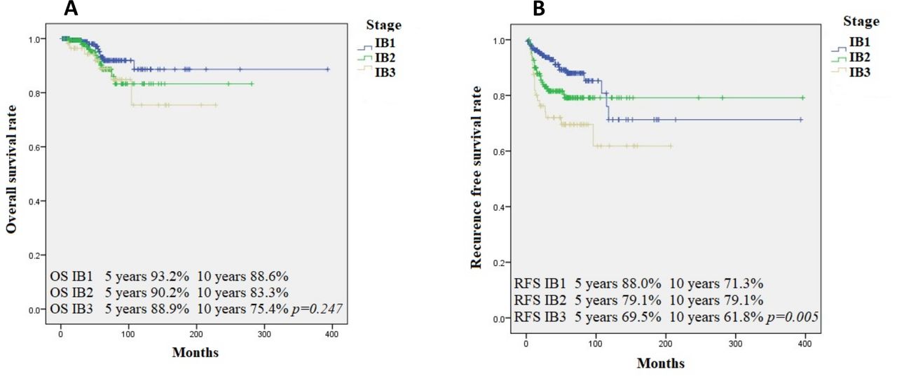
The 5-year overall survival rate was 91% and the 10-year overall survival rate was 84% for the entire cohort. The rates were 93% and 89%, respectively, for stage IB1 endocervical adenocarcinomas; 90% and 83%, respectively, for stage IB2 endocervical adenocarcinomas; and 89% and 75%, respectively, for stage IB3 endocervical adenocarcinomas (p=0.24). Kaplan–Meier curves are shown in Figure 1A. The 5-year and 10-year overall survival rates, as classified by the Silva system, were both 100% for Silva pattern A endocervical adenocarcinomas, both 92% for Silva pattern B endocervical adenocarcinomas, and 92% and 81%, respectively, for Silva pattern C endocervical adenocarcinomas (p=0.08). The 5-year overall survival rate was 93% and the 10-year overall survival rate was 87% in HPV-associated cases. In HPV-independent cases, the 5-year overall survival rate was 81% and the 10-year overall survival rate was 45% (p=0.007). The 5-year overall survival rate was 94% and the 10-year overall survival rate was 91% in lymphovascular invasion-negative cases. In lymphovascular invasion-positive cases, the 5-year overall survival rate was 88% and the 10-year overall survival rate was 76% (p=0.004).
(A) Kaplan–Meier curve for 5- and 10-year overal survival rates in patients with stage IB1, IB2, and IB3 endocervical adenocarcinomas. (B) Kaplan–Meier curve for 5- and 10-year recurrence-free survival rates in patients with stage IB1, IB2, and IB3 endocervical adenocarcinomas.
Survival analysis by Cox regression (univariate analysis) showed that factors significantly associated with overall survival included the following: HPV-independent status (p=0.01; HR 3.00; 95% CI 1.29 to 6.99), destructive invasive Silva patterns (p=0.047; HR 2.78; 95% CI 1.01 to 7.62), presence of lymphovascular invasion (p=0.006; HR 3.14; 95% CI 1.38 to 7.15), and presence of lymph node metastasis (p=0.002; HR 3.93; 95% CI 1.66 to 9.29) (Table 2). Survival analysis by Cox regression (univariate analysis) showed that the factor significantly associated with overall survival in stage IB1 cases was the presence of lymph node metastasis (p=0.011; HR 6.05; 95% CI 1.51 to 24.25) (Table 3).
Survival analysis by Cox regression (univariate analysis) in 464 FIGO stage IB endocervical adenocarcinomas
Survival analysis by Cox regression (univariate analysis) of parameters that influence overall survival in FIGO IB substages
Prognostic Biomarkers for Recurrence-free Survival
The overall 5-year recurrence-free survival rate was 82%, and the 10-year recurrence-free survival rate was 72% (p=0.045). The rates were 88% and 71%, respectively, for stage IB1 endocervical adenocarcinomas; 79% and 79%, respectively, for stage IB2 endocervical adenocarcinomas; and 70% and 62%, respectively, for stage IB3 endocervical adenocarcinomas (p=0.005, log rank) (Figure 1B). Stratified by Silva pattern, the 5-year recurrence-free survival rates were 92% (Silva pattern A), 84% (Silva pattern B), and 78% (Silva pattern C); the 10-year recurrence-free survival rates were 92% (Silva pattern A), 67% (Silva pattern B), and 65% (Silva pattern C) (p=0.04, log rank). The 5- and 10-year recurrence-free survival rates for HPV-associated cases were 84% and 72%, respectively. For HPV-independent cases, the rates were 63% and 42%, respectively (p=0.001). In lymphovascular invasion-negative cases, the 5-year and 10-year recurrence-free survival rates were 94% and 81%, respectively, while, in lymphovascular invasion-positive cases, the rates were 68% and 60%, respectively (p=0.00001).
Statistically significant factors associated with recurrence-free survival were FIGO stage (p=0.001; HR 1.67; 95% CI 1.22 to 2.30), HPV-independent status (p=0.001; HR 2.73; 95% CI 1.53 to 4.87), destructive invasive Silva patterns (p=0.015; HR 1.75; 95% CI 1.11 to 2.76), high histologic grade (p=0.002; HR 1.81; 95% CI 1.24 to 2.64), presence of lymphovascular invasion (p=0.00001; HR 5.00; 95% CI 2.71 to 9.22), absence of demonstrable precursor lesions (p=0.005; HR 2.13; 95% CI 1.25 to 3.63), and presence of lymph node metastasis (p=0.00001; HR 5.44; 95% CI 3.12 to 9.48) (Table 2).
On multivariate analysis for the entire cohort, factors that significantly influenced recurrence-free survival included HPV-independent status (p=0.05; HR 2.31; 95% CI 1.02 to 5.46), presence of lymphovascular invasion (p=0.011; HR 3.50; 95% CI 1.33 to 9.19), and the presence of lymph node metastasis (p=0.016; HR 2.66; 95% CI 1.20 to 5.90) (Table 4).
Multivariate analysis of factors that influence recurrence-free survival in 464 FIGO stage IB endocervical adenocarcinomas
On univariate analysis (Cox regression), statistically significant factors associated with recurrence-free survival in stage IB1 cases were the following: presence of lymphovascular invasion (p=0.001; HR 4.48; 95% CI 1.78 to 11.23), absence of precursor lesions (p=0.035; HR 2.71; 95% CI 1.07 to 6.84), HPV-independent status (p=0.046; HR 3.54; 95% CI 1.02 to 12.19), and presence of lymph node metastasis (p=0.008; HR 4.60; 95% CI 1.48 to 14.29) (Online supplemental table S1). On multivariate analysis, presence of lymphovascular invasion (p=0.02; HR 5.18; 95% CI 1.30 to 20.68) was the only parameter that influenced recurrence-free survival in stage IB1 cases (Online supplemental table S2). On multivariate analysis, high tumor grade and presence of lymphovascular invasion and lymph node metastasis were significantly associated with recurrence-free survival in stage IB2 cases. On multivariate analysis, lymphovascular invasion (p=0.043; HR 3.18; 95% CI 1.03 to 9.76) and lymph node metastasis (p=0.021; HR 2.97; 95% CI 1.17 to 7.54) were significantly associated with recurrence-free survival in stage IB2 cases. In stage IB3 cases, recurrence-free survival was influenced by HPV-independent status and the presence of lymph node metastasis on both univariate and multivariate analysis (Online supplemental tables S1 and S2).
Supplemental material
Supplemental material
When comparing sub-stages IB1 and IB2 on univariate analysis, significantly associated clinico-pathological parameters included the following: older age (p=0.044; OR 1.55; 95% CI 1.01 to 2.38), surgical treatment (p=0.02; OR 2.14; 95% CI 1.11 to 4.12), the presence of lymphovascular invasion (p=0.00001; OR 2.31; 95% CI 1.52 to 3.53), HPV-independent status (p=0.009; OR 2.61; 95% CI 1.24 to 5.46), and high histologic grade (p=0.024; OR 1.69; 95% CI 1.07 to 2.66) (Online supplemental table S3). On multivariate analysis (logistic regression), HPV-independent status and the presence of lymphovascular invasion were significantly associated (Online supplemental table S4).
Supplemental material
Supplemental material
When comparing sub-stages IB2 and 1B3 on univariate analysis, significantly associated clinico-pathological parameters included the following: presence of lymphovascular invasion (p=0.00001; OR 3.40; 95% CI 1.71 to 6.75), absence of precursor lesions (p=0.004; OR 2.63; 95% CI 1.35 to 5.12), HPV-independent status (p=0.001; OR 3.24; 95% CI 1.55 to 6.80), and histologic grade (p=0.021; OR 3.05; 95% CI 1.13 to 8.17) (Online supplemental table S3). On multivariate analysis (logistic regression), factors that remained statistically significant were presence of lymphovascular invasion (p=0.032; OR 2.83; 95% CI 1.09 to 7.36) and HPV-independent status (p=0.028; OR 2.97; 95% CI 1.12 to 7.87) (Online supplemental table S5).
Supplemental material
Discussion
Endocervical adenocarcinomas have been the subject of much recent study to reclassify this tumor based on etiology (HPV status) and to find alternative options (Silva system) for reporting depth of invasion—a parameter that is especially difficult to assess in endocervical adenocarcinomas—in order to better identify patients at risk for lymph node metastasis so that appropriate patients can have more precise tailored surgery.2 5
The 2018 FIGO staging system for cervical cancer does not differentiate cervical adenocarcinoma from squamous cell carcinoma. FIGO stage IA tumors are diagnosed on microscopic examination since these tumors are not clinically visible. Clinically visible lesions and those with a depth of invasion >5 mm are designated FIGO stage IB and then sub-staged according to largest tumor diameter (IB1, 6–20 mm; IB2, 21–40 mm; and IB3, >40 mm). However, 2018 FIGO stage IB endocervical adenocarcinomas represent a heterogeneous group of tumors, which pose clinical management challenges for gynecologic oncologists. Patients with FIGO stage IB1 endocervical adenocarcinoma are considered at lower risk for metastasis and death from disease if the tumor diameter is <20 mm, cervical stromal invasion is <50% of the cervical wall, and there are no suspicious lymph nodes on radiologic examination.6 For women who do not desire fertility preservation, the standard treatment for the majority of these patients is a radical (Type C1) hysterectomy with pelvic lymphadenectomy with or without sentinel lymph node mapping. For FIGO stage IB2 endocervical adenocarcinoma, primary treatment includes surgery (radical hysterectomy with pelvic lymphadenectomy with or without sentinel lymph node mapping) or chemoradiotherapy, depending on local resources and other factors.6 Patients with FIGO stage IB3 endocervical adenocarcinoma are considered at higher risk for metastasis and recurrence due to larger tumor size, deep cervical wall invasion, and the presence of lymphovascular invasion and lymph node metastasis. Depending on local resources and patient preference, treatment can include surgery (radical hysterectomy and pelvic lymphadenectomy) and pelvic radiation therapy with concurrent platinum-based chemotherapy.
Prognostic factors for endocervical adenocarcinomas are well known, but little is known about prognostic biomarkers influencing overall survival and recurrence-free survival for the newly defined FIGO 2018 stage IB adenocarcinoma sub-stages and, to the best of our knowledge, no studies regarding these parameters have been published.
We found that, in stage IB endocervical adenocarcinomas, recurrence-free survival was influenced by prognostic biomarkers such as HPV status, Silva pattern, and the presence of lymphovascular invasion and lymph node metastasis. Our data show that HPV status and the presence of lymphovascular invasion (two parameters that can be easily identified by pathologists on microscopic examination) are important local tumor prognosticators in and among stage IB sub-types. These biomarkers, independent of treatment, might influence recurrence-free survival and should be included in any future sub-staging modification of FIGO stage IB cases and in the treatment strategies of these cancers. Patients with HPV-independent adenocarcinoma (such as gastric type), particularly in association with lymphovascular invasion and lymph node metastasis, may need novel treatment and surgical oncologic strategies as they are less responsive to current chemotherapy and radiotherapy standard protocols. The current FIGO grading system has some value, and large size adenocarcinomas are mostly poorly differentiated. The Silva pattern may be most relevant for the smaller size lesions such as stage IB1 rather than IB2 and IB3 cases. The strength of the study lies in the large number of stage IB endocervical adenocarcinomas collected from various international institutions, which were reviewed by gynecologic pathology experts for all pathologic parameters including HPV status and Silva pattern of invasion. Moreover, all the identified important local tumor prognosticators in and among stage IB sub-types can be easily identified by pathologists on microscopic examination. A possible weakness of the study is the variation in surgical and/or oncologic treatment options from center to center. Another possible weakness is the mean follow-up of 4 years, and some cases had a shorter follow-up.
Conclusion
Tumor prognosticators should be included in future sub-staging modifications of FIGO stage IB endocervical adernocarcinomas as well as in treatment strategies for these tumors. As with stage IB cervical adenocarcinoma, further studies are needed to define the role of HPV status, the presence of lymphovascular invasion, and Silva pattern of invasion in FIGO stage IA cases.
Supplementary materials
Supplementary Data
This web only file has been produced by the BMJ Publishing Group from an electronic file supplied by the author(s) and has not been edited for content.
Footnotes
Twitter @Nouraalmadani
Contributors Conceptualization: SS, RAS, NRA-R. Data curation: all authors. Formal analysis: MB. Funding acquisition: RAS, KP, NRA-R. Investigation: SS, RAS, MB, NRA-R. Methodology: SS, RAS, NRA-R. Project administration: NRA-R. Resources; RAS, KP, NRA-R. Software: MB. Supervision: NRA-R. Validation: RAS, NRA-R. Visualization: SS. Roles/writing original draft: SS, RAS, NRA-R. Writing - review and editing: all authors.
Funding This research was funded in part through the National Institutes of Health/National Cancer Institute Cancer Center Support Grant P30 CA008748.
Competing interests Outside the submitted work, NRA-R reports grants from Stryker/Novadaq, Olympus, and GRAIL.
Patient consent for publication Not required.
Provenance and peer review Not commissioned; externally peer reviewed.
Data availability statement Data are available upon reasonable request. Please contact the corresponding author for data requests.
Supplemental material This content has been supplied by the author(s). It has not been vetted by BMJ Publishing Group Limited (BMJ) and may not have been peer-reviewed. Any opinions or recommendations discussed are solely those of the author(s) and are not endorsed by BMJ. BMJ disclaims all liability and responsibility arising from any reliance placed on the content. Where the content includes any translated material, BMJ does not warrant the accuracy and reliability of the translations (including but not limited to local regulations, clinical guidelines, terminology, drug names and drug dosages), and is not responsible for any error and/or omissions arising from translation and adaptation or otherwise.