Article Text

Abstract
Introduction There is limited evidence favoring the use of the sentinel lymph node technique in ovarian cancer, and no standardized approach has been studied. The objective of the present pilot study is to determine the feasibility of the sentinel lymph node technique by applying a clinical algorithm.
Methods Patients with confirmed ovarian cancer were included. 99mTc and indocyanine green were injected into the ovarian and infundubulo-pelvic ligament stump. A gamma probe and near-infrared fluorescence imaging were used for sentinel lymph node detection.
Results The sentinel lymph node technique was performed in ten patients with a detection rate in the pelvic and/or para-aortic region of 100%. The tracer distribution rates of sentinel lymph nodes in the pelvic and para-aortic regions were 87.5% and 70%, respectively.
Conclusion The detection of sentinel lymph nodes in early-stage ovarian cancer appears to be achievable. Based on these results, a clinical trial entitled SENTOV (SENtinel lymph node Technique in OVarian cancer) will be performed.
- ovarian cancer
- early stage
- sentinel lymph node
- staging
Statistics from Altmetric.com
HIGHLIGHTS
Detection of sentinel lymph nodes in early-stage ovarian cancer appears to be achievable.
The sentinel lymph node technique was performed in 10 patients with a detection of 100%.
The tracer distribution rates of sentinel lymph nodes in the pelvic and para-aortic regions were 87.5% and 70%.
Introduction
Epithelial ovarian cancer is diagnosed as a disease that is apparently limited to the pelvis (FIGO III) in up to 20% of cases.1 2 In this scenario, comprehensive surgical staging is recommended, including hysterectomy, unilateral/bilateral salpingo-oophorectomy, omentectomy and pelvic and para-aortic lymphadenectomy.3
Compared with other procedures, lymphadenectomy is the most complex, owing to the advanced surgical skills needed to achieve an appropriate and safe result.4 Nevertheless, it is associated with both intra-operative morbidity, such as vascular injury, and a significant post-operative morbidity, even when performed by experts.5 6
The justification for lymphadenectomy is explained by the possible presence of microscopic disease in the lymph nodes. In this case, it determines the prognosis and adjuvant systemic treatment needed,7 as the incidence of positive lymph nodes in patients with early-stage ovarian cancer is between 10–20%.8 Theoretically, the omission of an adequate lymphadenectomy in an early clinical stage may lead to the exclusion of adjuvant chemotherapy due to the under-diagnosis of lymph node metastasis. Despite these facts, no benefit of lymphadenectomy in terms of survival in patients with early epithelial ovarian cancer confined to the ovary has been demonstrated,9 and in cases of low-grade ovarian cancer, the incidence of lymph node metastasis has been reported recently to be rather low.10 11
The concept of sentinel lymph node consists of the detection of the first node of drainage from a tumor. The absence of metastasis predicts the nodal status of all lymph nodes in a certain anatomic region. The detection of a negative sentinel lymph node suggests that the remaining lymph nodes are not involved. Consequently, lymphadenectomy, and thus, the associated morbidity, can be avoided. The sentinel lymph node technique for assessing regional lymph node status has been widely studied and proven to be effective at replacing lymphadenectomy in breast and vulvar cancer.12 13 In other gynecologic cancers, such as cervical and uterine cancers, the sentinel node technique is currently halfway to standardization.14 15
Ovarian cancer in an early stage may suppose an ideal tumor case for application of the sentinel lymph node technique for the following reasons: lymphadenectomy is a difficult procedure associated with high morbidity and results in the absence of lymph node involvement in a high percentage of cases. Nevertheless, both technical difficulty and the risk of tumor dissemination associated with the injection of tracers into the ovarian cortex16 17 has limited the number of published studies.16–21 The objective of the present pilot study is to determine the feasibility of the sentinel lymph node technique in an apparent early-stage ovarian cancer by applying a clinical algorithm.
Methods
The study has been approved by the institutional review board and local ethics committee. Patients with suspicious or confirmed adnexal masses were assessed for eligibility. We prospectively included patients with apparent early-stage ovarian cancer who met the following inclusion criteria: >18 years of age, suspicious adnexal mass (unilateral or bilateral) at CT imaging or confirmed ovarian tumor after previous surgery (unilateral/bilateral salpingo-oophorectomy ± hysterectomy) and signed an informed consent form. All patients underwent total body CT scan to evaluate the extent of disease, including pre-operative assessment of nodal involvement.
The exclusion criteria were as follows: unsuspected tumor presence according to FIGO (International Federation of Gynecology and Obstetrics) stage III, previous vascular surgery (cava vein, aorta, iliac vessels), previous lymphadenectomy (pelvic or para-aortic), history of lymphoma, radiotherapy (pelvic or para-aortic fields), a benign result after frozen section in the case of a suspicious adnexal mass, and/or previous allergic reaction to colloids or indocyanine green.
For the detection of sentinel lymph nodes, two methods were used: 99mTc and indocyanine green. After the abdominal cavity was accessed, in cases of previously unconfirmed malignant histology, the suspicious ovarian tumor was removed. Unilateral or bilateral salpingo-oophorectomy was performed, but hysterectomy was performed only if it was necessary to avoid rupture of the tumor capsule. The surgical specimen was then submitted for frozen sectioning.22 In the case of malignancy, the sentinel lymph node technique was performed. We subperitoneally injected 0.2 mL of saline solution containing 37 mBq of 99mTc nanocolloid (Albu-res, Pharmaceutical Nycomed Amersham, Braunschweig, Germany). At the same time, 0.5 mL of indocyanine green (concentration 1.25 mg/mL) was injected. We used a 27 G needle at each injection point (online supplementary video).
Supplementary video
In cases of unilateral tumors and no previous hysterectomy, the injection points were at the infundibulo-pelvic and ovarian stumps. On the other hand, for bilateral tumors and no previous hysterectomy, the injection points were both infundibulo-pelvic and ovarian ligament stumps. If hysterectomy was performed before the sentinel lymph node technique, the injection was made only at one or both infundibulo-pelvic stumps in cases of unilateral or bilateral tumors, respectively.
After a minimum of 15 min, the point of injection and migration of the sentinel lymph nodes were checked with an intra-operative mobile gamma camera (Sentinellatm, Oncovision) for descriptive purposes only. Thirty minutes after the injection, the SLN procedure was started regardless of probe migration with the IMGC.
Guided by the acoustic signal of a gamma probe (Wprobe wireless gamma probe STD and LAP, Oncovision), we performed a minimum dissection looking for the hottest sentinel lymph node(s) in the pelvic/para-aortic region. We simultaneously used the Imagen1 HUB-OPAL1 (NIR/ICG system) (Karl Storz Endoscopy, GmbH, Mittelstrasse, Tuttlingen, Germany) to detect the sentinel lymph node(s) dyed with indocyanine green. Any lymph node with a remarkably higher count than the background was considered a sentinel lymph node and was harvested separately. All retrieved sentinel lymph nodes were classified according to the anatomical region in which they were located (Figure 1).
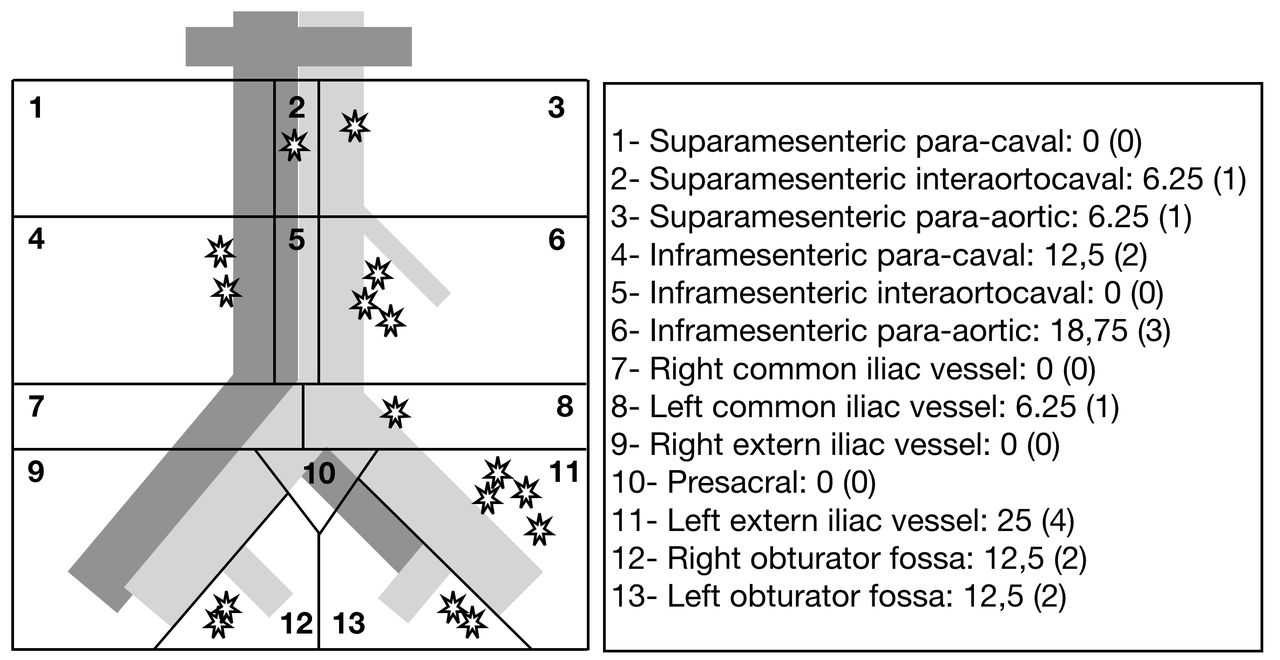
Distribution of sentinel lymph nodes.
After location and resection of sentinel lymph node(s), a systematic surgical staging was accomplished including: hysterectomy, contralateral adnexectomy if needed, pelvic and para-aortic lymphadenectomy, omentectomy, abdominal wash (performed after the initial abdominal access), and appendectomy in the case of mucinous tumors. Regarding the extent of the pelvic lymphadenectomy, dissection was performed within the origin of the external iliac vessels and continued caudally around them along the medial border of the psoas muscle until the deep inferior epigastric vessels were reached. The lateral limit of dissection was the fascia covering the psoas muscle, and the depth limit was the obturator nerve and vessels, including the lymphatic tissue from the obturator fossa. The medial margin of the lymphadenectomy was represented by an imaginary plane parallel to the umbilical artery. In addition, lymphatic tissue was cleared from the obturator fossa. Para-caval and para-aortic lymphadenectomy was performed by removing the inter-cavoaortic, pre-caval and pre-aortic nodal groups included between the renal vein (upper limit), common iliac vessel (lower limit), and both ureters (lateral limits), as well as the pre-sacral nodes.
Both sentinel lymph nodes and non-sentinel lymph nodes were processed according to a standard protocol for lymph node examination; these were cut into single sections or, in the case of a diameter >1 cm, into 2–3 sections and stained with hematoxylin-eosin before microscopy.
Results
Sixteen patients were considered for inclusion. Six of them were excluded for the following reasons: four cases due to a benign result of the intra-operative examination and two cases due to the inability to identify the target injection point because of severe adherence syndrome. Finally, the sentinel lymph node technique was performed in 10 patients.
The mean±SD age at diagnosis was 45±13.3 years, and the body mass index was 24.9±4.9 kg/m2. In cases where no previous surgery had been performed, malignancy was confirmed by intra-operative examination (Table 1). The approach was laparoscopy in three cases and laparotomy in the other seven cases due to the size of the tumor (Table 2). An intra-operative examination was performed in the two cases with no confirmation of malignancy, and the sentinel lymph node technique was not performed, as explained earlier. In one case, hysterectomy and bilateral salpingo-oophorectomy were needed during the intra-operative examination (due to the risk of tumor rupture). The mean±SD tumor size was 162±66 mm (range 75–250 mm) at diagnosis.
Baseline characteristics
Surgical data and sentinel lymph node results
In seven cases, 99mTc+indocyanine green was injected intra-operatively into the ovarian ligament stump. In the other two cases, it was not injected because a hysterectomy had been performed previously or during the intra-operative examination. Regarding the infundibulo-pelvic ligament stump, the injection was performed in all cases, unilateral injection in eight cases and bilateral injection in two other cases due to the involvement of both ovaries (Table 2). There were no adverse or clinically detectable pharmacologic effects in any of the ten subjects. Thirty minutes after injection, the point of injection and sentinel lymph node migration was assessed with an intra-operative mobile gamma camera (100% detection rate).
A sentinel lymph node was detected in the pelvic and/or para-aortic region in 10 (100%) and nine (90%) cases using the gamma probe and indocyanine green camera, respectively. The migration and tracer distribution rates of sentinel lymph nodes in the pelvic region were 87.5% (7/8) and 70% (7/10), respectively, in cases of para-aortic nodes. Mapping of the distribution of the sentinel lymph nodes detected is shown in Figure 1. The mean number of harvested sentinel lymph nodes was 2.55±1.6 (range 1–6): a mean of 1.86±1.35 (0–4) nodes in the pelvic field and 1.50±1.41 nodes (range 0–4) in the para-aortic region. The distribution of the retrieved sentinel lymph nodes is shown in Figure 1. The mean time from injection to the resection of sentinel lymph nodes was 54±31 min (range 25–120 min). Complete surgical staging was performed after the intra-operative examination and sentinel lymph node technique (if not previously performed) as follows: hysterectomy, contralateral salpingo-oophorectomy, systematic pelvic and para-aortic lymphadenectomy, omentectomy, and peritoneal cytology. The final histotypes were serous (six cases), endometrioid (one case) and clear cell (three cases). Of these, 20% (2/10) were classified as low grade and 80% (8/10) as high grade. The mean number of retrieved lymph nodes was 18±6 (range 12–30) and 20±9 (range 9–34) in the pelvic and para-aortic locations, respectively (Table 3). The final FIGO stage was IA in one case (10%), IC in five cases (50%), and IIA in one patient (10%). Two patients were finally upgraded to stage IIIA1 because of lymph node metastasis. One of them was found in one pelvic sentinel lymph node. The other was a positive para-aortic node in a patient in whom the sentinel lymph node was not identified at the para-aortic field because of the absence of migration. The other patient was upgraded to stage IIIA2 because of the presence of microscopic disease at the omentum.
Final histologic data
Discussion
To date, pelvic and para-aortic LND are the standard procedures in early ovarian cancer.3
Nevertheless, due to the lack of a benefit in terms of survival9 and the low incidence of microscopic lymph node metastasis8 10 11 associated with morbidity related to the LND,4 6 this issue remains controversial.
In this pilot study, we propose a clinical scheme for a sentinel lymph node technique in ovarian cancer and delineate its applicability. The approach was not a limitation, as the procedure was performed either by laparotomy or laparoscopy. Nevertheless, the presence of a severe adherence syndrome was the main hindrance to performing the technique. The use of 99mTc allowed the detection of a sentinel lymph node in at least one field (pelvic and/or para-aortic) in 100% of cases, and the use of indocyanine green was very useful for detection in most cases, with a detection rate of 90%.
Regarding each point of injection, migration and sentinel lymph node detection were easier to achieve in the pelvic versus para-aortic field, with a detection rate of 87.5% (7/8) and 70% (7/10), respectively.
One of the advantages of the proposed scheme is that the sentinel lymph node technique is only performed in cases of malignancy by intra-operative examination, restricting the use of 99mTc and indocyanine green. In contrast, the surgical time is extended by approximately 1 hour by the performance of the sentinel lymph node technique.
Two patients were upgraded to FIGO IIIA1. One presented metastasis at one of the sentinel lymph nodes harvested, and the rest of the pelvic and para-aortic nodes in this patient were negative. Hence, the negative predictive value of the SLN technique was 100%. In the other patient upgraded by lymph node metastasis, there was no migration-detection of sentinel lymph nodes at the para-aortic field, and a positive node was found among those retrieved after para-aortic LND.
Most of the previously published series16–21 explored the technique in patients with endometrial cancer or borderline ovarian tumors. Only some of the studies performed the technique in patients with apparent early-stage ovarian cancer: Kepple et al included six patients with epithelial ovarian cancer, Hassanzadeh et al included 11 patients, Buda et al included seven patients and Nyberg et al included four patients. However, none of them proposed a standardized approach to cases of suspected or confirmed ovarian cancer. Different points of injection have been proven. It has been described that tracers should be injected into both ovarian ligaments with a detection rate of between 90–100%. An injection near the hilum of the ovary has also been performed with a detection rate between 94–100%. Nevertheless, injection at this point is not feasible in the case of previously performed adnexectomy or if it is performed for the purpose of intra-operative examination. Injection into the cortex of the ovary has also been shown to have a worse detection rate (40–100%).
An assortment of different tracers has been used for SLN detection, such as CH40 (activated carbon particles), 99mTc albumin colloid, blue dye or indocyanine green, either alone or in combination, and 99mTc is the most commonly used. Nevertheless, a standardized dose has not yet been established. Based on previously published experiences, we delineated a protocol that could be adopted and prospectively integrated into clinical practice once confirmed by a clinical trial.
The role of single photon emission CT (SPECT) after the sentinel lymph node technique for the detection of residual lymph nodes has been studied by Speth et al.23 A discrepancy between the gammaprobe and SPECT was found. Nevertheless, most of the hotspots detected by SPECT after surgery were in the pelvic region, and were probably related to the injection sites. The presence of hotspots at the para-aortic area may be explained by the type of surgery performed (lymph node sampling).
Further studies are needed in order to delineate the tracer dose, point of injection and feasibility of the clinical algorithm for the sentinel lymph node technique in ovarian cancer. With this aim, a clinical trial entitled SENTOV (SENtinel lymph node Technique in OVarian cancer) will be performed in our center (EudraCT Number: 2017-003683-12).
Conclusions
In light of the findings of this pilot study, a clinical trial will be performed in our center to establish an evidence-based conclusion. The sentinel lymph node technique appears to be applicable in early-stage ovarian cancer, and the presence of severe adherence syndrome is the main concern when performing the technique. A future clinical trial entitled SENTOV (SENtinel lymph node Technique in Ovarian cancer) will be performed to delineate the tracer dose, point of injection and feasibility of the clinical algorithm for the sentinel lymph node technique in ovarian cancer (EudraCT number: 2017-003683-12).
Supplemental material
References
Footnotes
Funding The authors have not declared a specific grant for this research from any funding agency in the public, commercial or not-for-profit sectors.
Competing interests None declared.
Ethics approval Obtained.
Provenance and peer review Not commissioned, externally peer reviewed.