Article Text

Abstract
A European consensus conference on endometrial carcinoma was held in 2014 to produce multi-disciplinary evidence-based guidelines on selected questions. Given the large body of literature on the management of endometrial carcinoma published since 2014, the European Society of Gynaecological Oncology (ESGO), the European SocieTy for Radiotherapy and Oncology (ESTRO), and the European Society of Pathology (ESP) jointly decided to update these evidence-based guidelines and to cover new topics in order to improve the quality of care for women with endometrial carcinoma across Europe and worldwide.
- uterine cancer
- surgery
- radiation
- pathology
Statistics from Altmetric.com
Introduction
Endometrial carcinoma is the most common gynecological cancer in Europe, with a 5-year prevalence of 34.7% (445 805 cases).1 The estimated number of new endometrial carcinoma cases in Europe in 2018 was 121 578 with 29 638 deaths, and the incidence has been rising with aging and increased obesity of the population. The EUROCARE-5 study, published in 2015, reported a 5-year relative survival of 76% for European women diagnosed with endometrial carcinoma in 2000–2007, ranging from 72.9% in Eastern Europe to 83.2% in Northern Europe.2 The observed geographic difference might be partially attributable to tangible differences in the prevalence of endometrioid sub-types among regions. Furthermore, differences in patient characteristics and histopathologic features of the disease impact both on patient prognosis and the recommended treatment approach.
A consensus conference including representation from the European Society of Medical Oncology (ESMO), the European Society of Gynaecological Oncology (ESGO), and the European SocieTy for Radiotherapy and Oncology (ESTRO) was held in 2014 with the aim to produce multi-disciplinary evidence-based guidelines on 12 selected questions in order to complement the ESMO clinical practice guidelines previously published.3–6 ESGO, ESTRO, and the European Society of Pathology (ESP) jointly decided to update these evidence-based guidelines and, moreover, to cover new topics in order to provide comprehensive guidelines on all relevant issues of diagnosis and treatment in endometrial carcinoma in a multi-disciplinary setting. These guidelines are intended for use by gynecological oncologists, general gynecologists, surgeons, radiation oncologists, pathologists, medical and clinical oncologists, radiologists, general practitioners, palliative care teams, and allied health professionals.
Responsibilities
These guidelines are a statement of evidence and consensus of the authors regarding their views of currently accepted approaches for the management of patients with endometrial carcinoma. Any clinician applying or consulting these guidelines is expected to use independent medical judgment in the context of individual clinical circumstances to determine any patient’s care or treatment. These guidelines make no warranties of any kind regarding their content, use, or application, and the authors disclaim any responsibility for their application or use in any way.
Methods
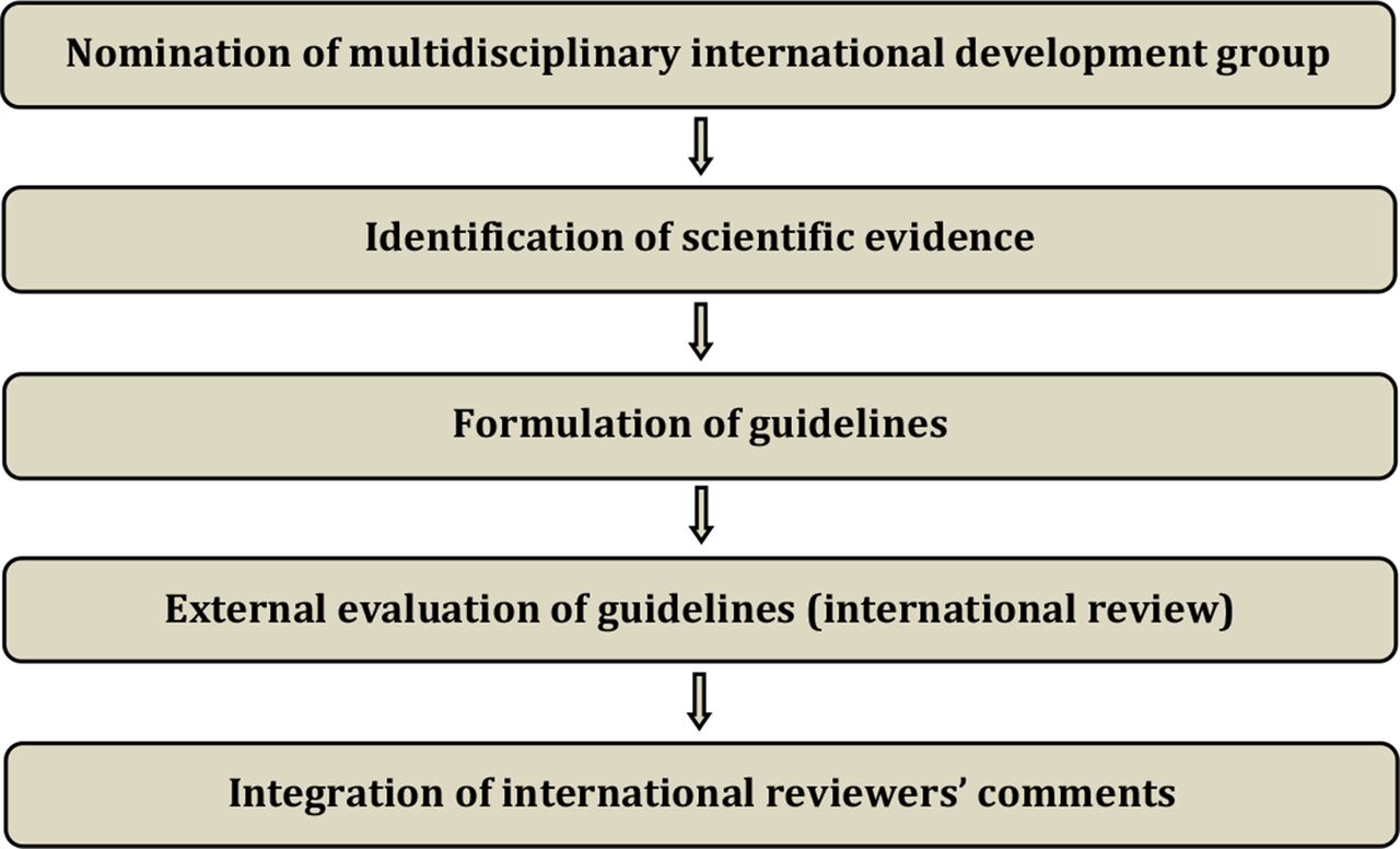
The guidelines were developed using a five-step process as defined by the ESGO Guideline Committee (see Figure 1). The strengths of the process include creation of a multi-disciplinary international development group, use of scientific evidence and international expert consensus to support the guidelines, and use of an international external review process (physicians and patients). This development process involved three meetings of the international development group chaired by Professor Nicole Concin (Medical University of Innsbruck, Innsbruck, Austria/Evangelische Kliniken Essen-Mitte, Essen, Germany, for ESGO), Professor Carien L Creutzberg (Leiden University Medical Center, Leiden, the Netherlands, for ESTRO), and Professor Xavier Matias-Guiu (Department of Pathology, Hospital Universitari Arnau de Vilanova and Hospital Universitari de Bellvitge, Irblleida, Idibell, Universities of Lleida and Barcelona, CIBERONC, Spain, for ESP).

Development process.
ESGO/ESTRO/ESP nominated practising clinicians who are involved in the management of patients with endometrial carcinoma and have demonstrated leadership in the clinical management of patients through research, administrative responsibilities, and/or committee membership to serve on the expert panel. The objective was to assemble a multi-disciplinary panel and it was therefore essential to include professionals from relevant disciplines (gynecological oncology and gynecology, medical, clinical and radiation oncology, pathology) to contribute to the validity and acceptability of the guidelines. To ensure that the statements were evidence based, the current literature was reviewed and critically appraised. A systematic literature review of relevant studies published between January 2014 and June 2019 was carried out using the MEDLINE database (see online supplemental appendix 1). The literature search was limited to publications in English. Priority was given to high-quality systematic reviews, meta-analyses, and randomized controlled trials, but studies of lower levels of evidence were also evaluated. The search strategy excluded editorials, letters, and in vitro studies. The reference list of each identified article was also reviewed for other potentially relevant articles.
Supplemental material
The development group developed guidelines for all the topics. The guidelines were retained if they were supported by a sufficiently high level of scientific evidence and/or when a large consensus among experts was obtained. An adapted version of the 'Infectious Diseases Society of America-United States Public Health Service Grading System' was used to define the level of evidence and grade of recommendation for each of the recommendations7 (see Table 1). In the absence of any clear scientific evidence, judgment was based on the professional experience and consensus of the development group.
Levels of evidence and grades of recommendations
ESGO/ESTRO/ESP established a large multi-disciplinary panel of practicing clinicians who provide care to patients with endometrial carcinoma to act as independent expert reviewers for the guidelines developed. These reviewers were selected according to their expertise, had to be still involved in clinical practice, and were from different European and non-European countries to ensure global perspective. Patients with endometrial carcinoma were also included. These independent reviewers were asked to evaluate each recommendation according to its relevance and feasibility in clinical practice (only physicians), so that comprehensive quantitative and qualitative evaluations of the guidelines were completed. Patients were asked to evaluate qualitatively each recommendation (according to their experience, personal perceptions, etc). Evaluations of the external reviewers (n=191) were pooled and discussed by the international development group before finalising the guidelines. The list of the 191 external reviewers is available in online supplemental appendix 2.
Supplemental material
General recommendations
Planning of staging and treatment should be made on a multi-disciplinary basis (generally at a tumor board meeting, composed according to local guidelines) and based on the comprehensive and precise knowledge of prognostic and predictive factors for outcome, morbidity, and quality of life (V, A).
Patients should be carefully counseled about the suggested diagnostic and treatment plan and potential alternatives, including risks and benefits of all options (V, A).
Treatment should be undertaken in a specialized center by a dedicated team of specialists in the diagnosis and management of gynecological cancers, especially in high-risk and/or advanced stage disease (V, A).
Identification and surveillance of women with a pathogenic germline variant in a Lynch syndrome-associated gene
Approximately 3% of all endometrial carcinomas and about 10% of mismatch repair deficient (MMRd)/microsatellite unstable endometrial carcinomas are causally related to germline mutations of one of the MMR genes MLH1, PMS2, MSH2 and MSH6.8 Testing for MMR status/microsatellite instability (MSI) in endometrial carcinoma patients has been shown to be relevant for four reasons: (1) diagnostic, as MMRd/MSI is considered a marker for endometrioid-type endometrial carcinoma; (2) pre-screening to identify patients at higher risk for having Lynch syndrome; (3) prognostic, as identified by The Cancer Genome Atlas (TCGA, see below for molecular classification); and (4) predictive for potential utility of immune checkpoint inhibitor therapy. The International Society of Gynecological Pathology (ISGyP) has recommended testing for MMR status/MSI in all endometrial carcinoma samples, irrespective of age.9 This has also been recommended in other society statements and recommendations, such as the Manchester International Consensus Group recommendations, whenever resources are available.10
The preferred approach (widely available and cost-effective) to identifying patients with a higher chance of having Lynch syndrome is by MMR-immunohistochemistry (IHC) on well preserved tumor tissue. MMR-IHC is a reliable method to assess MMR status, and in addition provides information on the altered gene/protein. ISGyP guidelines therefore recommend MMR-IHC as the preferred test.9 MMR-IHC consists of the assessment of the expression of four MMR proteins (MLH1, PMS2, MSH6, and MSH2). A simplified two-antibody (PMS2 and MSH6) approach has been proposed as a cost-effective alternative.11–13 This procedure still requires performing MLH1 and MSH2 IHC in cases with any abnormal staining of PMS2 and/or MSH6. Molecular analyses for the microsatellite status (MSI-test) are an alternative, but are more laborious, require non-neoplastic tissue, are more expensive, and do not provide information on the gene affected. For optimal pre-selection of patients at risk for having Lynch syndrome, both approaches require the analysis of MLH1 promoter methylation status in cases with loss of MLH1/PMS2 expression. Testing for MMRd by IHC or MSI by PCR-based methods does not allow direct identification of patients with Lynch syndrome since MMRd/MSI is frequently due to sporadic events such as bi-allelic somatic mutations or hypermethylation. In the absence of hypermethylation, referral to genetic counseling is recommended to evaluate the presence of a germline mutation. When familial history is highly suspicious of Lynch syndrome, genetic counseling is recommended independent of the MMR status.
The cumulative incidences for cancer depend on the specific mutation in women with Lynch sydrome. For endometrial carcinoma, the cumulative incidences at 70 years are 34%, 51%, 49%, and 24% for MLH1, MSH2, MSH6, and PMS2 mutation carriers, respectively, and for ovarian cancer 11%, 15%, 0%, and 0%, respectively.14 Furthermore, the age of cancer onset in Lynch syndrome varies among specific mutated genes and types of mutations.15 Ryan et al suggest gynecological surveillance to be appropriate from age 30 years for those with MSH2 mutations, from age 35 years for those with nontruncating MLH1 mutations, and from age 40 years for those with MSH6 and truncating MLH1 mutations. Women with heterozygous PMS2 mutations do not warrant gynecological surveillance because their absolute risk of gynecological cancer is very low. As part of a retrospective study, Lachiewicz et al reported a risk of any occult malignancy during prophylactic surgery for women with Lynch syndrome or hereditary non-polyposis colorectal cancer to be up to 17%.16 Thus, these patients should be counseled about the risk of detection of gynecological cancer at prophylactic surgery.
Recommendations
To identify patients with Lynch syndrome and triage for germline mutational analysis, MMR IHC (plus analysis of MLH1 promotor methylation status in case of immunohistochemical loss of MLH1/PMS2 expression) or MSI tests should be performed in all endometrial carcinomas, irrespective of histologic subtype of the tumor (III, B).
Endometrial carcinoma patients identified as having an increased risk of Lynch syndrome should be offered genetic counseling (III, B).
Surveillance for endometrial carcinoma in Lynch syndrome mutation carriers should in general start at the age of 35 years; however, individual factors need to be taken into consideration (tailored surveillance programs). The decision on the starting age of surveillance should integrate knowledge on the specific mutation and history of onset of events in the family (IV, B).
Surveillance of the endometrium by annual transvaginal ultrasound (TVUS) and annual or biennial biopsy until hysterectomy should be considered in all Lynch syndrome mutation carriers (IV, B).
Hysterectomy and bilateral salpingo-oophorectomy to prevent endometrial and ovarian cancer should be performed at the completion of childbearing and preferably before the age of 40 years. All the pros and cons of prophylactic surgery must be discussed including the risk of occult gynecological cancer detection at prophylactic surgery. Estrogen replacement therapy should be suggested if bilateral salpingo-oophorectomy is performed in pre-menopausal women (IV, B).
Molecular markers for endometrial carcinoma diagnosis and as determinants for treatment decisions
Different types of endometrial carcinoma have specific histological and molecular features, precursor lesions and natural histories. Conventional pathologic analysis remains an important tool for tumor stratification, but suffers from inter-observer variation. Different groups have applied a diagnostic algorithm using three immunohistochemical markers (p53, MSH6 and PMS2) and one molecular test (mutation analysis of the exonuclease domain of POLE) to identify prognostic groups analogous to the TCGA molecular-based classification.17–21 The feasibility of this approach was confirmed by a large number of publications that have all consistently reported prognostic relevance particularly in high-grade and high-risk tumors in several independent cohorts and prospective clinical trials.22 To apply this molecular classification, all these diagnostic tests need to be performed. Performing one of the surrogate marker tests in isolation is insufficient, as a combination of positive tests can occur in approximatively 5% of all carcinomas. The diagnostic algorithm to classify these so-called 'multiple classifiers' has been described recently.23 24 In addition, endometrial carcinoma should only be classified as POLE-mutated (POLEmut) when pathogenic variants of POLE are identified in the gene’s exonuclease domain.25 26
This surrogate marker approach to the molecular-based classification has been demonstrated to be prognostically informative in low-, intermediate-, and high-risk endometrial carcinoma. Smaller studies showed that the molecular classification is also applicable to non-endometrioid tumors including serous, clear cell, undifferentiated carcinomas, and uterine carcinosarcomas. For adjuvant treatment recommendations, the molecular classification seems to be particularly relevant in the context of high-grade and/or high-risk endometrial carcinomas. Application of the molecular classification in high-grade and/or high-risk endometrial carcinomas shows that there is a group of patients with an excellent prognosis—that is, the POLEmut tumors—and a group with a poor prognosis—that is, the p53-abnormal (p53abn) tumors. Endometrial carcinomas with MMRd or non-specific molecular profile (NSMP) have an intermediate prognosis. However, the molecular surrogate is not perfect. Immunohistochemical demonstration of p53abn is a good but not perfect surrogate of TP53 mutation. Furthermore, a small proportion of high copy number tumors do not show TP53 mutations. To minimize these limitations, an integrated analysis combining traditional pathologic and molecular results seems ideal. In low-risk endometrioid carcinomas, the molecular classification may not be required.27 28
The proposed molecular classification of endometrial carcinoma is clinically feasible using a limited set of diagnostic tests. Using this novel classification is encouraged. All diagnostic tests should be performed in conjunction due to the occurrence of 'double classifiers'.23 Clinical management may be particularly impacted by the molecular classification in scenarios where adjuvant chemotherapy is considered (high-grade/high-risk disease). Thus, these cases should be prioritized when there is a lack of sufficient resources to perform this classification on all endometrial carcinomas. If molecular classification tools are not available, endometrial carcinoma classification should be based on traditional pathologic features. There is still room for other biomarkers that may be potentially useful in the big group of low-grade endometrioid carcinoma with NSMP, such as L1CAM expression or mutations in CTNNB1.29–32
Recommendations
Molecular classification is encouraged in all endometrial carcinomas, especially high-grade tumors (IV, B).
POLE mutation analysis may be omitted in low-risk and intermediate-risk endometrial carcinoma with low-grade histology (IV, C).
Definition of prognostic risk groups integrating molecular markers
There is overwhelming evidence that traditional pathologic features, such as histopathologic type, grade, myometrial invasion, and lymphovascular space invasion (LVSI), are important in assessing prognosis, as recommended in the ISGyP guidelines.9 Histopathologic typing should be performed according to the WHO Classification of Tumors (5th edition).33 A binary International Federation of Gynecology and Obstetrics (FIGO) grading is recommended, which considers grade 1 and grade 2 carcinomas as low-grade and grade 3 carcinomas as high-grade. For the assessment of myometrial invasion, account needs to be taken of the endo-myometrial junction which is undulating.34 Focal LVSI is defined by the presence of a single focus around the tumor, substantial LVSI as multifocal or diffuse arrangement of LVSI or the presence of tumor cells in five or more lymphovascular spaces. The molecular classification adds another layer of information to the conventional morphologic features and therefore should be integrated in the pathologic report.
Recommendations
Histopathologic type, grade, myometrial invasion, and LVSI (no/focal/substantial) should be recorded in all patients with endometrial carcinoma (V, A).
The definition of prognostic risk groups is presented in Table 2 for both situations when molecular classification is known or unknown.
Definition of prognostic risk groups
Pre- and intra-operative work-up
Risk group allocation on biopsy according to the WHO Classification of Tumors (5th edition) and FIGO grading of endometrial carcinoma is required for adequate planning of therapy.33 Histopathologic grade has prognostic relevance. A modified binary FIGO grading is recommended lumping together grade 1 and grade 2 endometrioid carcinomas as low-grade and grade 3 as high-grade.
Magnetic resonance imaging (MRI) techniques are highly specific in the assessment of deep myometrial invasion, cervical stromal involvement, and lymph node metastasis.35–82 The diagnostic performance of TVUS and MRI for detecting myometrial invasion in endometrial carcinoma are quite similar.39 44 56 83–88 Of note, pre-operative ultrasound assessment of deep myometrial and cervical stromal invasion in endometrial carcinoma is best performed by an expert sonographer as, compared with gynecologists, they show a greater degree of agreement with histopathology and greater inter-observer reproducibility.84 Positron emission tomography (PET) scanning has an excellent specificity for the pre-operative assessment of lymph node metastases in patients with endometrial carcinoma. Its moderate sensitivity for detecting lymph node metastases during preo-perative staging probably reflects the need for a sufficient number of neoplastic cells to induce 18F-fluoro-2-deoxy-D-glucose hypermetabolism.89–100 The usefulness of maximal standardized uptake value in classifying patients into pre-defined risk groups is limited.101 A pre-operative CT scan has a clinical utility in patients with endometrial carcinoma in detecting metastatic disease.102 103
Frozen section of endometrial biopsy material is obsolete. Myometrial invasion should not be assessed by frozen section because of poor reproducibility and agreement with definitive paraffin sections. Since sentinel node biopsy is increasingly used, the need for intra-operative assessment of myometrial invasion has become less important. Moreover, some of the biomarkers that have been proposed require optimal management of the surgical specimen with high quality pre-analytical issues such as appropriate fixation conditions. Performing frozen sections can lead to incorrect control of pre-analytical conditions, sometimes even leading to incorrect assessment of LVSI due to artifactual displacement of tumor cells into vascular spaces during processing. In addition, the freezing of tissue before fixation and further processing interferes with an optimal pre-analytical procedure required for standardized histopathologic diagnosis.
Recommendations
Histopathologic tumor type and grade in endometrial biopsy is required (IV, A).
Pre-operative mandatory work-up includes: family history; general assessment and inventory of co-morbidities; geriatric assessment, if appropriate; clinical examination, including pelvic examination; expert transvaginal or transrectal ultrasound or pelvic MRI (IV, C).
Depending on clinical and pathologic risk, additional imaging modalities (thoracic, abdominal and pelvic CT scan, MRI, PET scan, or ultrasound) should be considered to assess ovarian, nodal, peritoneal, and other sites of metastatic disease (IV, C).
Intra-operative frozen section is not encouraged for myometrial invasion assessment because of poor reproducibility and interference with adequate pathologic processing (IV, A).
Early stage disease
Surgical management of apparent stage I/II endometrial carcinomas
Minimally invasive approach
Two randomized prospective studies comparing minimally invasive with open surgeries showed similar survival with quicker recovery with the minimally invasive approach.104 105 More recently, pooled analyses of randomized prospective studies including, notably, these two studies and multiple retrospective and prospective studies also support the use of minimally invasive surgery for patients including those with high-risk endometrial carcinoma.106–171
Recommendations
Minimally invasive surgery is the preferred surgical approach, including patients with high-risk endometrial carcinoma (I, A).
Any intra-peritoneal tumor spillage, including tumor rupture or morcellation (including in a bag), should be avoided (III, B).
If vaginal extraction risks uterine rupture, other measures should be taken (eg, mini-laparotomy, use of endobag) (III, B).
Tumors with metastases outside the uterus and cervix (excluding lymph node metastases) are relative contra-indications for minimally invasive surgery (III, B).
Standard surgical procedures
In a randomized controlled trial comparing modified radical (Piver–Rutledge class II) hysterectomy to the standard extrafascial (Piver–Rutledge class I) or simple total hysterectomy in stage I endometrial carcinoma, Signorelli et al showed no differences in locoregional control and survival.172 The high risk of microscopic omental metastases in stage I serous and undifferentiated endometrial carcinoma and in carcinosarcoma suggests that omentectomy should be part of staging surgery in these patients.173 The low rate of omental metastases in apparent clinical stage I endometrioid and clear cell carcinoma does not justify the procedure.174 Although the risk of having occult (microscopic) omental metastases in carcinosarcoma is around 6%, staging omentectomy in these women is suggested. Identification of these cases will allow inclusion of patients with advanced stage disease into clinical trials.175 Positive peritoneal cytology correlates with poor prognostic factors and poor survival; however, it is not part of FIGO staging and unclear if this should influence treatment decisions.176–178
Recommendations
Standard surgery is total hysterectomy with bilateral salpingo-oophorectomy without vaginal cuff resection (II, A).
Staging infracolic omentectomy should be performed in clinical stage I serous endometrial carcinoma, carcinosarcoma, and undifferentiated carcinoma. It can be omitted in clear cell and endometrioid carcinoma in stage I disease (IV, B).
Surgical re-staging can be considered in previously incompletely staged patients with high– intermediate-risk/high-risk disease if the outcome might have an implication for adjuvant treatment strategy (IV, B).
Lymph node staging
Sentinel node biopsy has been introduced as an alternative to lymph node dissection for lymph node staging and, if done according to state-of-art principles, a negative sentinel node is accepted to confirm pN0. Multiple studies, including prospective cohort ones, confirmed high sensitivity of sentinel lymph node status for lymph node staging in patients with early-stage endometrial carcinoma and support the impact of sentinel lymph node biopsy on surgical management and indications for adjuvant therapies.179–241 More intensive pathologic assessment of sentinel lymph node (sentinel lymph node ultrastaging) supports the detection of small metastases which could be missed by standard evaluation.214 232 Sentinel lymph node biopsy without dissection of other pelvic lymph nodes is associated with subtantially lower risk of post-operative morbidity, especially lower leg lymphedema.242 In a large group of patients with low-risk (myometrial invasion <50%, low-grade) endometrial carcinoma with sentinel lymph node biopsy, lymph node involvement was found in 6% of patients, half of them identified by pathologic ultrastaging.243 Patients with tumors without myometrial invasion did not have any positive sentinel lymph nodes. Four prospective cohort trials have shown high sensitivity to detect pelvic lymph node metastases and a high negative predictive value by applying a sentinel lymph node algorithm in high-risk/high-grade endometrial carcinomas in the hands of experienced surgeons. 181 182 237 244 Recently, a randomized controlled trial highlighted that the use of indocyanine green instead of methylene blue dye resulted in a significant increase in sentinel lymph node detection rates per hemipelvis in women with endometrial carcinoma undergoing minimally invasive surgery.245 Retrospective studies showed a similar prognosis for patients after full lymphadenectomy and sentinel lymph node biopsy only.179 201 220 High bilateral pelvic sentinel lymph node detection can be achieved when the tracer is injected into the cervix.180 246 A higher sentinel lymph node detection rate has been reported using near-infrared fluorescence in comparison to other techniques.247 A worse prognosis is associated with the presence of nodal micrometastases, especially in patients who do not receive adjuvant treatment.248 There is no evidence that the presence of isolated tumor cells (ITCs) has an impact on prognosis, and similar to other tumor sites, the stage would be pN0(i+). If pelvic lymph node involvement is reported either by sentinel lymph node frozen section or by the final pathology, para-aortic staging can be considered, either by imaging (with all limitations of the imaging modalities) or by surgery. It should be noted that, based on data from two large randomized trials, lymph node staging does not have a therapeutic value but is done to assess the extent of disease and to provide information for adjuvant treatment decisions.249 250 Frozen section on specimens regarded as sentinel lymph nodes can confirm the presence of lymph nodes and macrometastases but should not replace adequate pathologic processing and ultrastaging.
Recommendations
Sentinel lymph node biopsy can be considered for staging purposes in patients with low-risk/intermediate-risk disease. It can be omitted in cases without myometrial invasion. Systematic lymphadenectomy is not recommended in this group (II, A).
Surgical lymph node staging should be performed in patients with high–intermediate-risk/high-risk disease. Sentinel lymph node biopsy is an acceptable alternative to systematic lymphadenectomy for lymph node staging in stage I/II (III, B).
If sentinel lymph node biopsy is performed (II, A):
Indocyanine green with cervical injection is the preferred detection technique.
Tracer re-injection is an option if sentinel lymph node is not visualized upfront.
Side-specific systematic lymphadenectomy should be performed in high–intermediate-risk/high-risk patients if sentinel lymph node is not detected on either pelvic side.
Pathologic ultrastaging of sentinel lymph nodes is recommended.
When a systematic lymphadenectomy is performed, pelvic and para-aortic infrarenal lymph node dissection is suggested (III, B).
Presence of both macrometastases and micrometastases (<2 mm, pN1(mi)) is regarded as a metastatic involvement (IV, C).
The prognostic significance of ITCs, pN0(i+), is still uncertain (IV, C).
If pelvic lymph node involvement is found intra-operatively, further systematic pelvic lymph node dissection should be omitted. However, debulking of enlarged lymph nodes and para-aortic staging can be considered (IV, B).
Option for ovarian preservation and salpingectomy in stage I/II
A meta-analysis showed that there was no significant difference in overall survival between patients treated with ovarian preservation and bilateral salpingo-oophorectomy.251 A similar result was achieved in young and pre-menopausal women. Disease-free survival of patients whose ovaries were preserved was slightly compromised, but this was not statistically significant. Ovarian preservation can be cautiously considered in specific clinical situations when treating young and pre-menopausal women with early stage endometrial carcinoma because it is not associated with a significant adverse impact on survival.252–254 Salpingectomy during hysterectomy is recommended to decrease the risk of high-grade serous ovarian carcinoma.255 Ovarian preservation is not recommended in patients with cancer family history involving ovarian cancer risk (eg, BRCA mutation, Lynch syndrome, etc), but oocyte cryopreservation might be considered.256
Recommendations
Ovarian preservation can be considered in pre-menopausal patients aged <45 years with low-grade endometrioid endometrial carcinoma with myometrial invasion <50% and no obvious ovarian or other extra-uterine disease (IV, A).
In cases of ovarian preservation, salpingectomy is recommended (IV, B).
Ovarian preservation is not recommended for patients with cancer family history involving ovarian cancer risk (eg, BRCA mutation, Lynch syndrome, etc) (IV, B).
Radicality of surgery for clinical stage II
Radicality of hysterectomy (simple vs modified radical hysterectomy (type II)) in stage I–III endometrial carcinoma has no impact on local recurrence rate, disease-free survival, and overall survival. In a meta-analysis enrolling 2866 patients with stage II endometrial carcinoma, radical hysterectomy did not show a significant survival benefit for either overall survival or progression-free survival compared with simple hysterectomy.257 The result remained consistent after it was adjusted for the possible impact of adjuvant radiotherapy.
Recommendations
Total hysterectomy with bilateral salpingo-oophorectomy and lymph node staging is the surgical standard of care in patients with stage II endometrial carcinoma (IV, B).
More extensive procedures should only be performed if required to achieve free surgical margins (IV, B).
Medically unfit patients
It is rare for patients to be unfit for surgery, but medical co-morbidities, which increasingly include morbid obesity, can preclude surgery due to high operative and peri-operative risks. Ideally, assessment should be undertaken in a center with specialist anesthetic experience in managing these high-risk patients. Definitive radiotherapy with brachytherapy, external beam radiation therapy (EBRT) or the combination of both modalities can be considered.258–262
Recommendations
Medical contra-indications to the standard surgical management by minimally invasive surgery are rare. Vaginal hysterectomy, with bilateral salpingo-oophorectomy if feasible, can be considered in patients unfit for the recommended standard surgical therapy (IV, C).
Definitive radiotherapy can be considered for primary tumors where surgery is contra-indicated for medical reasons:
The combination of EBRT and brachytherapy should be used for high-grade tumors and/or deep myometrial invasion (II, B).
For low-grade tumors, brachytherapy alone can be considered (II, B).
In medically unfit patients unsuitable for curative surgery or radiotherapy, systemic treatment (including hormonal therapy) can be considered (IV, B).
Fertility preservation
Work-up for fertility preservation treatments
Fertility-sparing treatments should be considered in patients with atypical hyperplasia/endometrioid intra-epithelial neoplasia (AH/EIN) or grade 1 endometrioid carcinoma without myometrial invasion.263–269 There are very few published data on patients with stage IA grade 2 endometrioid carcinoma without myometrial invasion who received fertility-sparing treatment with combined oral medroxyprogesterone acetate/levonorgestrel intrauterine system.270 Although results are encouraging, this treatment should only be considered by experienced gynecological oncologists using well-defined protocols with detailed patient information and close follow-up.
Hysteroscopic biopsy is suggested, based on its higher agreement with the final diagnosis compared with dilatation and curettage.271 272 Although hysteroscopy seems to be associated with a higher rate of positive peritoneal cytology, it seems not to have a negative impact on survival.273 Expert vaginal ultrasound examination can be used instead of pelvic MRI. Its high diagnostic performance allows the detection of myometrial invasion and cervical stromal invasion with respect to final pathologic examination. Ultrasound should be performed by an expert sonographer (a practitioner who spends a significant part of her/his time undertaking ultrasound examinations in gynecology and gynecologic oncology and has fulfilled the minimum training requirements for level 3 following the recommendations of the European Federation of Societies for Ultrasound in Medicine and Biology).274
There is currently a lack of high-quality evidence regarding the correlation between weight loss and reduction of risk of recurrence/increased survival in patients with endometrial carcinoma, especially with respect to fertility-sparing treatment.275 Diabetes mellitus does not seem to affect the outcome of conservative treatment in women with AH/EIN or early endometrial carcinoma.276 Conversely, the use of metformin seems associated with an improvement in overall survival for patients with endometrial carcinoma and a reduced risk of cancer relapse.277 In addition, metformin is associated with improvement in the overall survival of patients with endometrial carcinoma with diabetes.
Recommendations
Patients who are candidates for fertility-preserving treatment must be referred to specialized centers. Fertility-sparing treatment should be considered only in patients with AH/EIN or grade 1 endometrioid endometrial carcinoma without myometrial invasion and without genetic risk factors (V, A).
In these patients, endometrial biopsy, preferably through hysteroscopy, must be performed (III, A).
AH/EIN or grade 1 endometrioid endometrial carcinoma must be confirmed/diagnosed by a pathologist experienced in gynecological pathology (V, A).
Radiologic imaging to assess the extension of the disease must be performed. An expert ultrasound examination can substitute pelvic MRI scan (III, B).
Patients must be informed that fertility-sparing treatment is not a standard treatment. Only patients who strongly desire to preserve fertility should be treated conservatively. Patients must be willing to accept close follow-up and be informed of the need for future hysterectomy in case of failure of treatment and/or after pregnancies (V, A).
Management and follow-up for fertility preservation
To date, there are no available randomized controlled trials comparing different methods of conservative treatment in women with AH/EIN or presumed stage IA grade 1 endometrioid carcinoma. Existing data suggest that patients who received hysteroscopic resection followed by progestin therapy achieve the highest complete remission rate compared with other existing fertility-preserving treatments.263–269 278–295 Intrauterine progestin therapy such as levonorgestrel-releasing intrauterine system combined with gonadotropin-release hormone receptor agonist/progestin have a satisfactory pregnancy rate and low recurrence rate. Patients who received oral progestin only might be more likely to recur and have more systemic adverse effects.
Recommendations
All patients should be evaluated before and after the fertility-sparing treatment at a fertility clinic (IV, C).
Hysteroscopic resection prior to progestin therapy can be considered (III, B).
Medroxyprogesterone acetate (400–600 mg/day) or megestrol acetate (160–320 mg/day) is the recommended treatment. Treatment with levonorgestrel intrauterine device in combination with oral progestins with or without gonadotropin-releasing hormone analogs can also be considered (IV, B).
In order to assess response, hysteroscopic guided biopsy and imaging at 3–4 and 6 months must be performed. If no response is achieved after 6 months, standard surgical treatment is recommended (IV, B).
Continuous hormonal treatment should be considered in responders who wish to delay pregnancy (IV, B).
Strict surveillance is recommended every 6 months with TVUS and physical examination. During follow-up, hysteroscopic and endometrial biopsy should be performed only in case of abnormal uterine bleeding or atypical ultrasound findings (IV, B).
Fertility-sparing treatment can be considered for intrauterine recurrences only in highly selected cases under strict surveillance (IV, C).
Hysterectomy and bilateral salpingo-oophorectomy is recommended after childbearing due to a high recurrence rate. Preservation of the ovaries can be considered depending on age and genetic risk factors (IV, B).
Synchronous presentation of low-grade endometrioid endometrial and ovarian carcinomas
Adnexal involvement by endometrial carcinoma is currently a parameter important in FIGO staging and has an impact on overall survival rate.296 It was shown that patients with simultaneous involvement of the endometrium and ovary by low-grade endometrioid carcinoma had a favorable outcome. This suggested that they were synchronous primary tumors rather than metastatic sites. Several criteria have been used in the past to distinguish between endometrial carcinoma with ovarian metastasis and synchronous primary tumors.297 298 However, these were not easy to apply.
Recent studies have shown that, for low-grade endometrioid carcinomas, there is a clonal relationship between endometrial and ovarian carcinomas in the vast majority of cases, indicating that the carcinoma arises in the endometrium and extends secondarily to the ovary.299 300 In the most recent edition of WHO (2020) it is mentioned that patients with clonally related low-grade endometrioid carcinomas should be managed without adjuvant treatment (as if they were two independent primaries) when fulfilling the following criteria: (1) low-grade endometrioid morphology, (2) no more than superficial myometrial invasion, (3) absence of LVSI, and (4) absence of additional metastases.33 301
Recommendation
If all WHO 2020 criteria mentioned above are met and the ovarian carcinoma is pT1a, no adjuvant treatment is recommended (III, B).
Adjuvant treatment
Adjuvant treatment recommendations for endometrial carcinoma strongly depend on the prognostic risk group (see Table 2 for definitions of the prognostic risk groups with and without known molecular classification).
Low risk
For patients with low-risk endometrial carcinoma, no adjuvant treatment is recommended based on data from multiple randomized trials.302–305 For patients with stage I–II POLEmut endometrial carcinomas, no adjuvant treatment seems justifiable based on the data from independent series showing very few recurrences and also in cases of observation.21 25 For stage III patients, however, there are only indirect data to support this, as all cases with advanced disease had adjuvant treatment. In the molecular analysis of the PORTEC-3 trial of high-risk endometrial carcinoma, those with POLEmut endometrioid carcinoma had an excellent outcome in both arms.22 However, both trial arms included EBRT. Prospective registration (preferably in national or international studies) of POLEmut endometrial carcinoma cases with treatment and outcome data is strongly recommended.
Recommendations
For patients with low-risk endometrial carcinoma, no adjuvant treatment is recommended (I, A).
When molecular classification is known:
For patients with endometrial carcinoma stage I–II, low-risk based on pathogenic POLE-mutation, omission of adjuvant treatment should be considered (III, A).
For the rare patients with endometrial carcinoma stage III–IVA and pathogenic POLE-mutation, there are no outcome data with the omission of the adjuvant treatment. Prospective registration is recommended (IV, C).
Intermediate risk
Adjuvant brachytherapy provides excellent vaginal control and high survival rates, similar to those after adjuvant EBRT in this intermediate-risk population, as shown in large randomized trials, particularly the PORTEC-2 trial and Swedish trial.306–314 It was also shown that only the small minority of patients with higher risk based on substantial LVSI, p53abn, or L1CAM overexpression had a slightly higher risk of pelvic recurrence with vaginal brachytherapy than those who had EBRT. Therefore, the intermediate-risk category only includes those with none or only focal LVSI and no p53abn. In a Danish population study it was confirmed that the risk of locoregional relapse was higher (about 14%) with omission of vaginal brachytherapy, but that overall survival was not different due to treatment of relapse.315 Therefore, no adjuvant treatment is an option in this group, especially for patients aged <60 years who have a lower risk of relapse.
MMRd and, especially, NSMP cancers form the majority of endometrioid carcinomas and have an intermediate prognosis, in between POLEmut (excellent prognosis) and p53abn carcinomas (unfavorable prognosis). Findings of prior large randomized trials in high–intermediate-risk endometrial carcinoma are therefore mainly applicable to MMRd and NSMP endometrioid carcinomas in this intermediate-risk category.
It has to be stressed that p53abn carcinomas restricted to a polyp or without myometrial invasion were not included in the randomized trials and the value of chemotherapy and of EBRT are uncertain. Since the studies mentioned above did not include and/or did not address non-endometrioid (and/or p53abn) carcinomas without myometrial invasion, there are very few specific available data on the best treatment for stage IA non-endometrioid carcinomas (serous, clear cell, undifferentiated carcinoma, carcinosarcoma, mixed) without myometrial invasion. Some case series and a recent analysis using the US National Cancer Data Base suggest that adjuvant chemotherapy (with or without vaginal brachytherapy) might improve survival, while other reports showed good outcomes with vaginal brachytherapy only.306 Therefore, these carcinomas have been grouped in the intermediate-risk category and adjuvant therapy should be discussed on a case-by-case basis until more prospective data are available.
Recommendations
Adjuvant brachytherapy can be recommended to decrease vaginal recurrence (I, A).
Omission of adjuvant brachytherapy can be considered (III, C), especially for patients aged <60 years (II, A).
When molecular classification is known, POLEmut and p53abn with myometrial invasion have specific recommendations (see respective recommendations for low- and high-risk).
For p53abn carcinomas restricted to a polyp or without myometrial invasion, adjuvant therapy is generally not recommended (III, C).
High–intermediate risk (pN0 after lymph node staging)
The definition of high–intermediate risk has changed in comparison with the ESMO-ESGO-ESTRO consensus conference. In the current prognostic risk group classification (see Table 2), stage IA endometrioid carcinomas are only included if there is substantial LVSI.3–5 This high–intermediate-risk group also includes stage IB low-grade endometrioid with substantial LVSI, and stage IB high-grade endometrioid carcinomas regardless of LVSI, and stage II endometrioid carcinomas. In view of the higher risk of recurrence in this newly classified group (even with negative nodes), adjuvant brachytherapy can be recommended to decrease vaginal recurrence. In the case of substantial LVSI and/or stage II, EBRT can be considered as it has been shown to reduce the risk of pelvic and para-aortic nodal relapse.316
In two older randomized controlled trials317 318 there was no difference between adjuvant chemotherapy alone and EBRT alone in recurrence-free and overall survival. In the NSGO/EORTC trial and the PORTEC-3 trials, the combination of chemotherapy and radiotherapy seemed to provide better recurrence-free and overall survival outcomes respectively compared with radiotherapy alone.319 320 The GOG-249 trial did not find benefit in recurrence-free or overall survival from three cycles of chemotherapy with brachytherapy compared with EBRT alone.316 Molecular analysis of PORTEC-3 trial tissues suggested no benefit of chemotherapy for MMRd carcinomas.320 321 Omission of adjuvant treatment is an option and this should be considered only when close follow-up is guaranteed to ensure detection and prompt treatment of recurrence at an early stage.
Recommendations
Adjuvant brachytherapy can be recommended to decrease vaginal recurrence (II, B).
EBRT can be considered for substantial LVSI and for stage II (I, B).
Adjuvant chemotherapy can be considered, especially for high-grade and/or substantial LVSI (II, C).
Omission of any adjuvant treatment is an option (IV, C).
When molecular classification is known, POLEmut and p53abn have specific recommendations (see respective recommendations for low- and high-risk).
High–intermediate risk cN0/pNx (lymph node staging not performed)
In view of the recent randomized trials GOG-249 (for stage I and II endometrial carcinomas with high-risk factors or serous or clear cell histology), the PORTEC-3 trial and the older GOG-99 trial, adjuvant EBRT is recommended in case of substantial LVSI or stage II.302 316 319 320 322 Additional chemotherapy can be considered, especially for high-grade carcinomas, based on the PORTEC-3 trial, but the question remains whether the benefit outweighs the toxicity for stage I–II endometrioid carcinomas, and multi-disciplinary shared decision-making is needed.320 Molecular analysis of PORTEC-3 trial tissues suggested no benefit of chemotherapy for MMRd carcinomas.320 321 Adjuvant brachytherapy alone can be considered for LVSI negative cases and for stage II grade 1 disease.
Recommendations
Adjuvant EBRT is recommended, especially for substantial LVSI and/or for stage II (I, A).
Additional adjuvant chemotherapy can be considered, especially for high-grade and/or substantial LVSI (II, B).
Adjuvant brachytherapy alone can be considered for high-grade LVSI negative and for stage II grade 1 endometrioid carcinomas (II, B).
When molecular classification is known, POLEmut and p53abn have specific recommendations (see respective recommendations for low- and high-risk).
High risk
The risk category changes also have a substantial impact on this category. Some carcinomas designated as high risk in the ESMO-ESGO-ESTRO consensus conference are not included anymore in the high-risk sub-group in these ESGO-ESTRO-ESP guidelines.3–5 High-risk carcinomas are now either stage III–IVA without residual disease or stage I–IVA p53abn or non-endometrioid carcinomas without residual disease with myometrial invasion (for specifics see Table 2).
In 2019 the updated results of the PORTEC-3 trial, with a longer median follow-up of 72 months and with 75% of participants having reached 5 years of follow-up, were published.323 In this trial comparing combined chemotherapy and radiotherapy (two cycles of cisplatin during radiotherapy followed by four cycles of carboplatin-paclitaxel) with radiotherapy alone, a statistically significant 5% overall survival benefit at 5 years and a 7% failure-free survival benefit was seen in the combined therapy group compared with radiotherapy alone. The greatest overall survival difference was seen in stage III carcinomas and in serous carcinomas regardless of stage. The GOG-258 trial compared the same chemotherapy-radiotherapy schedule used in PORTEC-3 with six cycles of carboplatin-paclitaxel chemotherapy alone and found overlapping relapse-free and overall survival rates.324 However, the chemotherapy alone arm had significantly higher rates of pelvic and peri-aortic nodal relapse. Therefore, chemotherapy alone is an alternative option based on the GOG-258 results for stage III–IV disease. The final analysis of the GOG-249 trial highlighted that a post-operative adjuvant strategy of vaginal cuff brachytherapy followed by three cycles of paclitaxel and carboplatin chemotherapy did not significantly increase 5-year recurrence-free survival or 5-year overall survival compared with pelvic radiotherapy.325 Vaginal and distant recurrence rates were similar between arms. However, pelvic or para-aortic nodal recurrences were significantly less common with pelvic radiotherapy. The older pooled analysis of the NSGO-EORTC and MANGO-ILIADE trials used sequential chemotherapy and radiotherapy (either sequence) and reported significantly longer recurrence-free survival compared with radiotherapy alone.319 Multiple retrospective studies indicated a survival benefit in patients with advanced stage endometrial carcinoma treated with post-operative combined treatment including radiotherapy and chemotherapy, delivered by either the sandwich or sequential method, compared with radiotherapy alone or chemotherapy alone.326–344
The benefit of added chemotherapy is unclear for patients with stage I–II clear cell carcinomas. These have often been included with serous as 'non-endometrioid carcinomas'. Of note, in the PORTEC-3 trial it was specifically in those with serous histology that a significant benefit of added chemotherapy was seen.323 However, this was not observed in the NSGO-EORTC and MANGO-ILEADE trials. Extended field radiotherapy is used in the case of involved para-aortic nodes or involvement of high common iliac nodes, both with or without chemotherapy. The combination of extended field radiotherapy with chemotherapy using modern intensity-modulated radiation therapy/volumetric modulated arc therapy (IMRT/VMAT) techniques has been shown feasible in the PORTEC-3 and GOG-258 trials. An additional brachytherapy boost can be considered, especially for substantial LVSI, endocervical stromal invasion, and/or stage IIIB–IIIC.
MMRd and NSMP carcinomas are included in the high-risk category if stage III–IVA with no residual disease. The p53abn carcinomas can be of endometrioid, serous, undifferentiated, and clear cell histologic type, but all consistently show a poor outcome and should therefore be regarded as high risk. Based on the current data, it is more difficult to draw conclusions regarding carcinosarcomas and undifferentiated carcinomas that are NSMP endometrial carcinomas due to the lack of large series. For clear cell carcinomas, the available data suggest some prognostic information may lie in the molecular classification. About 40–50% of clear cell carcinomas are p53abn. While serous carcinomas in the PORTEC-3 trial had an unfavorable outcome and significant benefit of added adjuvant chemotherapy, those with clear cell carcinomas seemed to have an outcome similar to high-grade carcinomas in general and were more favorable if not p53abn.321 323 The findings of the randomized trials for endometrioid carcinomas cited above are therefore largely applicable to stage III MMRd and NSMP carcinomas and to stage I–III p53abn carcinomas. This was also seen in the molecular analysis of the PORTEC-3 trial, which showed a statistically significant survival advantage for p53abn carcinomas with combined therapy for stage I–III. In contrast, POLEmut carcinomas had almost no recurrences in both arms. There was no clear benefit of added chemotherapy for MMRd, while the NSMP carcinomas had some benefit of added chemotherapy especially in case of stage III. Prospective evaluation of the molecular characteristics in randomized trials is highly recommended.
Recommendations
EBRT with concurrent and adjuvant chemotherapy (I, A) or alternatively sequential chemotherapy and radiotherapy is recommended (I, B).
Chemotherapy alone is an alternative option (I, B).
Carcinosarcomas should be treated as high-risk carcinomas (not as sarcomas) (IV, B).
When the molecular classification is known, p53abn carcinomas without myometrial invasion and POLEmut have specific recommendations (see respective recommendations for low- and intermediate-risk) (III, C).
Advanced disease
Surgery for clinically overt stage III and IV disease
In stage III and IV endometrial carcinoma (including carcinosarcoma), maximal cytoreduction should be considered only if macroscopic complete resection is feasible with acceptable morbidity.345–350 Surgery should be performed in a specialized center. Pre-operative complete staging and multi-disciplinary discussion within a tumor board should be performed. Suspicious enlarged lymph nodes should be resected if complete resection is possible.351 352 A full systematic pelvic and para-aortic lymphadenectomy of non-suspicious lymph nodes should not be performed because there is no evidence of a therapeutic impact. If upfront surgery is not feasible or acceptable and therefore primary systemic therapy is given, delayed surgery can be considered in case of a meaningful response to chemotherapy.353–360
Recommendations
In stage III and IV endometrial carcinoma (including carcinosarcoma), surgical tumor debulking including enlarged lymph nodes should be considered when complete macroscopic resection is feasible with an acceptable morbidity and quality of life profile, following full pre-operative staging and discussion by a multi-disciplinary team (IV, B).
Primary systemic therapy should be used if upfront surgery is not feasible or acceptable (IV, A).
In cases of a good response to systemic therapy, delayed surgery can be considered (IV, C).
Only enlarged lymph nodes should be resected. Systematic lymphadenectomy is not recommended (IV, B).
Unresectable primary tumor due to local extent of disease
For patients presenting with unresectable locally advanced disease and no evidence of multiple distant metastases, treatment options include definitive radiotherapy or neoadjuvant chemotherapy followed by surgery or definitive radiotherapy, depending on response.261 354–356 361 Definitive radiotherapy comprises EBRT to the pelvis followed by image-guided brachytherapy. Concurrent chemotherapy may be considered to enhance the radiation effect. Brachytherapy should boost sites of macroscopic disease in the uterus, parametrium, or vagina using the ESTRO principles. Adjuvant chemotherapy should also be considered following primary local treatment (surgery or radiotherapy) to reduce the risk of distant metastases.
Recommendations
For unresectable tumors, multi-disciplinary team discussion should consider definitive radiotherapy with EBRT and intrauterine brachytherapy, or neoadjuvant chemotherapy prior to surgical resection or definitive radiotherapy, depending on response (IV, C).
Image-guided brachytherapy is recommended to boost intrauterine, parametrial, or vaginal disease (IV, A).
Chemotherapy should be considered after definitive radiotherapy (IV, B).
Residual pelvic or para-aortic lymph nodes following surgery
Residual lymph node disease can be treated with EBRT using an integrated or sequential boost to escalate the nodal dose. An IMRT technique reduces the risk of toxicity to surrounding tissue.362 Adjuvant chemotherapy reduces the risk of distant metastases for patients with lymph node involvement.320 323 324
Recommendations
Residual lymph node disease should be treated with a combination of chemotherapy and EBRT (III, B) or chemotherapy alone (IV, B).
EBRT should be delivered to pelvis and para-aortic nodes with dose escalation to involved nodes using an integrated or sequential boost (IV, B).
Residual pelvic disease (positive resection margin, vaginal disease, pelvic side wall disease)
Patients with residual pelvic disease following surgery have a high risk of both local and distant recurrence. Radiotherapy can achieve long-term local control while chemotherapy reduces the risk of distant metastases. An individualized approach with either (chemo)-radiotherapy to the pelvis followed by chemotherapy or adjuvant chemotherapy followed by radiotherapy to the pelvis±para-aortic nodes should be considered.
Recommendation
An individualized approach with either radiotherapy or chemotherapy or a combination of both modalities should be considered by a multi-disciplinary team (V, B).
Recurrent disease
Radiotherapy naïve patients
Treatment of patients with recurrent endometrial carcinoma involves a multi-disciplinary approach with surgery, radiotherapy, and/or systemic therapy depending on the fitness and wishes of the patient, the tumor dissemination patterns, and prior treatment. A decision about surgery needs to take account of patient morbidity and wishes, available non-surgical treatments, and resources. The interval between primary treatment and recurrence should also be taken into consideration. Patients with recurrent disease, including resectable peritoneal and lymph node relapse, should be considered for surgery only if it is anticipated that complete resection of macroscopic disease can be achieved with a reasonable morbidity profile.363–369 The extent of the operation will depend on the degree of tumor dissemination pattern.
Locoregional recurrence of endometrial carcinoma is rare. With the advent of modern image-guided radiation therapy, including IMRT and image-guided adaptive brachytherapy, radiotherapy has become the treatment of choice in previously non-irradiated patients with isolated vaginal recurrence or locoregional recurrence.363 364 370–379 Consideration should be given to remove solitary easily accessible vaginal relapses, for better local symptom control prior to radiotherapy.
Recommendations
Patients with recurrent disease (including peritoneal and lymph node relapse) should be considered for surgery only if it is anticipated that complete removal of macroscopic disease can be achieved with acceptable morbidity. Systemic and/or radiation therapy should be considered post-operatively depending on the extent and pattern of relapse and the amount of residual disease (IV, C).
In selected cases, palliative surgery can be performed to alleviate symptoms (eg, bleeding, fistula, bowel obstruction) (IV, B).
For locoregional recurrence, the preferred primary therapy should be EBRT±chemotherapy with brachytherapy (IV, A).
An easily accessable superficial vaginal tumor can be resected vaginally prior to radiotherapy (IV, C).
For vaginal cuff recurrence:
Pelvic EBRT+intracavitary (±interstitial) image-guided brachytherapy is recommended (IV, A).
In case of superficial tumors, intracavitary brachytherapy alone can be considered (IV, A).
Systemic treatment can be considered before or after radiotherapy (IV, C).
Radiotherapy pre-treated patients with locoregional recurrence
In patients who have previously received EBRT±brachytherapy, radical surgery with the intention of complete resection with clear margins should be considered in specialized centers after ruling out metastatic disease with modern imaging. Pelvic exenteration may be considered for central local relapse.349 380 381 Otherwise, further radiation should be considered as radical therapy with or without systemic therapy. Interstitial brachytherapy (low-dose rate or high-dose rate) as the sole modality of treatment or combined with EBRT can result in high local control over 1–5 years.374 ,375 382 383 Other techniques like permanent seed implant or post-operative electron irradiation, protons and stereotactic body radiotherapy may be recommended in highly selected patients.384–386 The appropriate dose for each case needs to be individualized. Some low-dose rate data suggest improved outcomes with doses >50 Gy. The high-dose rate data are more varied, suggesting improved local control with doses >40 Gy. In general, a longer time interval between the first and second course of radiation as well as recurrences <2–4 cm tend to have improved outcomes. Multi-disciplinary management is critical to develop individualized plans and to clearly communicate potential side effects and expected treatment outcomes.
Recommendations
In patients with a history of previous radiation, radical surgery, including exenteration, should be considered when the intention is complete resection with clear margins (IV, B).
Additional options to consider include intra-operative electron radiation therapy or other forms of radiation therapy (IV, C).
If surgery is not feasible, radical re-irradiation options include stereotactic body radiotherapy targeting the recurrence, permanent seed implants, or proton therapy. In selected cases, limited volume re-irradiation with EBRT and brachytherapy boost may be an option (especially if longer interval from the first irradiation) (IV, C).
In patients who only had previous brachytherapy, EBRT+brachytherapy boost is recommended (IV, C).
In patients where re-irradiation with ERBT is not an option, image-guided interstitial brachytherapy only is recommended (may improve outcome) (IV, C).
Oligometastatic recurrent disease
Oligometastases is a disease concept that is defined by a state of limited metastatic tumors for which local ablative therapy could be curative. It refers in general to cancer patients with 1–5 metastases or recurrences.387–389 In recent years the concept of oligometastatic relapse has evolved and has led to a change in the approach to treatment. A prolonged disease-free interval and perhaps even cure may be achieved in some situations where the primary cancer site (if still present) is controlled and metastatic sites are ablated (surgically or with radiation).390–393 Multi-disciplinary management is critical to develop individualized plans and to communicate potential side effects and expected treatment outcomes. The additional benefit of chemotherapy is uncertain.
Recommendations
Patients with oligometastatic disease should be considered for radical local therapy (IV, B).
Treatment options include (IV, B):
Surgery
Radiation therapy including stereotactic radiotherapy
Local ablating techniques
The additional benefit of chemotherapy is uncertain (IV, B).
Systemic treatment for recurrent disease
Hormonal treatment results in a response rate of up to 55% in advanced/recurrent endometrial carcinoma.394 Low-grade, slowly progressing, hormone receptor-positive tumors appear to gain the greatest benefit from treatment; however, clinical benefit has also been observed in patients with hormone receptor-negative tumors.395 Progestogens are generally recommended.395 Alternative options include aromatases inhibitors, tamoxifen, and fulvestrant. In the PARAGON trial a response rate of 7% and a clinical benefit rate of 44% was reported with anastrazole in a cohort of 82 patients with recurrent, receptor positive, endometrial carcinoma.396 A single-arm phase II trial demonstrated a high response rate and clinical benefit rate with the combination of letrozole and everolimus.397 Confirmation of hormone receptor status by biopsy should be considered at the time of recurrence because of a potential change in hormone receptor expression between primary tumor and recurrence. In patients undergoing hormonal therapy, the risk of thrombo-embolic events needs to be taken into account. Prophylaxis with low molecular weight heparin should be considered in patients at high risk for thrombosis and be given according to local guidelines. There are no universally agreed recommendations to predict a response to hormonal therapy in endometrial carcinoma based on estrogen and progesterone receptor immunohistochemical status. Some of the following should be taken into account: (1) a wide range of hormonal agents are used, including medroxyprogesterone acetate and synthetic progestational agents, luteinizing hormone releasing hormone antagonists, tamoxifen, and new generations of selective estrogen receptor modulators; each has a different molecular action and may therefore have different activity; (2) receptor-negative status is not an absolute contra-indication to hormone treatment; (3) in some reports, response rates to various hormonal treatments for patients with endometrial carcinoma are higher for those with progesterone receptor expression; (4) the methodology for assessing and scoring hormone receptor expression in endometrial carcinoma is variable in the reported series; (5) assessment of estrogen and progesterone receptor status in the primary tumor may not reflect the status in the recurrent or metastatic tumor and thus a biopsy of recurrent or metastatic carcinomas for hormone receptor analysis may be helpful; (6) from a pragmatic viewpoint, it seems reasonable to interpret a carcinoma as receptor positive when immunoreactivity for estrogen receptor or progesterone receptors is found in more than 1% of carcinoma cells, until stronger validated scientific evidence is provided.
The combination of carboplatin and paclitaxel is the standard chemotherapy treatment of advanced/recurrent endometrial carcinoma based on a randomized phase 3 trial comparing carboplatin-paclitaxel versus carboplatin-paclitaxel-anthracyclines that reported overlapping progression-free survival and overall survival between the two arms but an increased toxicity for the triple combination.398 No standard treatment has been identified as second-line therapy; a response rate of about 10–15% has been seen among all the available treatment options. Thus, enrollment of patients in clinical trials is strongly encouraged. Weekly paclitaxel and anthracyclines (including pegylated liposomal doxorubicin when available) are considered to be active drugs. The re-introduction of carboplatin may be considered after a prolonged interval from the last platinum treatment, based on the results of a single-center retrospective series in patients treated with a median platinum-free interval of 25 (8–79) months. A response rate of 50% and median progression-free and median overall survival of 10 and 27 months, respectively, was reported after platinum re-challenge.399
Several anti PD-1 and anti PD-L1 checkpoint inhibitors have been shown to have activity in endometrial carcinoma and thus far pembrolizumab has been approved by the Food and Drug Administration (FDA) based on the results of a phase 2 single arm trial for the treatment of MSI-high (MSI-H)/MMRd solid tumors that have progressed on conventional therapy.400 401 The combination of intravenous pembrolizumab and lenvatinib, an oral multi-receptor tyrosine kinase inhibitor, received FDA approval in October 2019 for the second-line systemic therapy of microsatellite-stable (ie, non-MSI-H/MMRd) endometrial carcinoma based on the results of a phase 2 single-arm trial reporting 36% response rate in this population, including significant activity in those with serous carcinoma.402 403 No phase 3 randomized data are yet available.
Approximately 30% of uterine serous carcinomas show HER2/neu over-expression. A small randomized phase 2 trial of paclitaxel and carboplatin with or without trastuzumab in HER2/neu positive disease showed a 4.6 month increase in median progression-free survival.404 Anti-angiogenic agents and PI3kinase/mTor and MEK inhibitors also have demonstrated activity but secure evidence of benefit is inconclusive due to the limited sample size of the trials, inconsistency of results, and the low therapeutic index of the drugs, suggesting further investigations in well-designed and properly powered molecularly driven randomized trials are warranted.405–416
Recommendations
Hormone therapy is the preferred front-line systemic therapy for patients with low-grade carcinomas without rapidly progressive disease (II, A).
Progestogens (medroxyprogesterone acetate 200 (–300) mg and megestrol acetate 160 mg) are recommended (III, A).
Alternative options for hormonal therapies include aromatases inhibitors, tamoxifen, fulvestrant (III, C).
The standard chemotherapy treatment is carboplatin AUC 5–6 + paclitaxel 175 mg/m2 every 21 days for six cycles (I, A).
There is no standard of care for second-line chemotherapy. Doxorubicin and paclitaxel are considered the most active therapies (IV, C).
In patients with a long platinum-free interval, re-introduction of platinum can be considered (IV, C).
Anti-PD1-based immune therapy with pembrolizumab could be considered for second-line therapy of MSI/MMRd carcinomas. The combination of pembrolizumab and the multi-tyrosine-kinase inhibitor lenvatinib could be considered for second-line treatment of microsatellite-stable carcinomas (III, B). However, its use may be limited due to regulatory approvals or reimbursement in different countries. Clinical trial participation should be offered to all patients with relapse disease (V, B).
Palliative radiotherapy
Historically, radiotherapy has been an efficient treatment to palliate bleeding and pain from pelvic disease or systemic metastases. This results in rapid pain relief and temporary cessation of bleeding in the majority of patients.417
Recommendations
Radiotherapy is indicated for palliation of symptoms related to pelvic or systemic disease (IV, A).
Hypofractionated small volume EBRT can be used for treating primary disease in patients not fit for radical treatment (IV, B).
Principles of radiotherapy
The following sections present the general principles, the principles of adjuvant radiotherapy, of definitive treatment, and of radiotherapy for recurrent disease.258–261 307 362 372 377 418–423
General principles
State-of-art techniques and radiotherapy dose are chosen based on clinical findings, pathology, and patient factors including co-morbidities. For complex treatments or rare cases, referral to a specialized center is recommended. Prospective assessment of toxicity is recommended. Patients should have counseling on pelvic care and general and sexual rehabilitation whenever appropriate.
Adjuvant radiotherapy
Radiotherapy should preferably commence within 6 (–8) weeks of surgery or be scheduled in relation to chemotherapy.
EBRT
IMRT/VMAT techniques are recommended because the more conformal dose distribution increases normal tissue-sparing compared with a four-field conventional or 3D-conformal plan.424 The clinical target volume (CTV) includes the pelvic nodes (external iliac, internal iliac, obturator, distal common iliac), parametria, and upper vagina. The upper common iliac and sub-aortic pre-sacral lymph nodes are included when there is cervical stromal involvement and/or pelvic lymph node involvement. The lymph node target volume may be extended to include the aortic bifurcation or para-aortic nodes, up to or just above the level of the renal vessels, depending on the location and number of positive lymph nodes, site of sentinel lymph nodes, and whether there is extrauterine primary tumor involvement. The CTV should be individualized when there is a positive resection margin, pelvic peritoneal disease, or vaginal involvement. Treatment with a comfortably full bladder reduces the volume of irradiated small bowel and bladder. The planning target volume (PTV) should account for potential internal motion, depending on the method of verification used during the course of treatment. Image-guided radiotherapy by repeated volumetric imaging with cone beam CT (and use of so-called library of plans or plan of the day techniques) may enable the use of smaller CTV-PTV margins to reduce normal tissue toxicity. The prescription dose is commonly 45–50.4 Gy in 25–28 fractions over 5–6 weeks. An integrated or sequential EBRT boost is given to residual lymph node disease, sites of extracapsular nodal spread, and positive lateral resection margins with a total dose of 55–60 Gy EQD210 for microscopic residual disease, or up to 66 Gy for macroscopic/bulky disease. Concurrent and adjuvant chemotherapy may be considered for stage III disease, serous histology and/or recurrent disease.
Vaginal brachytherapy
Vaginal examination is undertaken to ensure the vaginal cuff is healed and to assess the size and shape of the vagina to guide applicator selection. Usually a vaginal cylinder is used but other applicators can be used, depending on patient anatomy. The target volume is individually determined and is usually the upper third of the vagina to a depth of 5 mm (both superiorly and halfway along the active length). The high-dose rate brachytherapy dose is most commonly 21–24 Gy in 3–4 fractions to 0.5 cm from the applicator surface, or 8–11 Gy in 2–3 fractions when given as a boost following EBRT. A higher dose is required for treatment of residual disease or positive margins. Pulsed-dose rate brachytherapy can be used following EBRT to boost macroscopic residual disease witha dose of 15–25 Gy. The treatment planning options are to use a standard library plan for each applicator size and treatment length or to use image-guided adaptive brachytherapy. In institutions where image-guided adaptive brachytherapy is applied, imaging of the applicator with CT scan or MRI evaluates whether the applicator is in close apposition to the vaginal mucosa and close to organs at risk. This allows verification and calculation of cumulative doses, especially if vaginal brachytherapy is used as a boost after EBRT. Image-guided adaptive brachytherapy is strongly recommended when there is residual vaginal disease following surgery using similar principles to treatment for recurrent disease.
Definitive treatment
Definitive radiotherapy with EBRT, brachytherapy, or a combination of both is indicated for primary tumors where surgery is contra-indicated for medical reasons. If patients are medically unfit for surgery, consider whether a long course of EBRT would be tolerated or, if not, a more hypofractionated approach could be used. Intrauterine brachytherapy as a sole treatment modality is used for low-grade early stage disease whereas the combination of EBRT and intra-cavitary brachytherapy is recommended for high-grade tumors and/or deep myometrial invasion. Specialist anesthetic review may be required to assess suitability for brachytherapy or whether brachytherapy could be applied with local anesthesia only. More advanced inoperable disease is treated with a combination of pelvic EBRT and intrauterine brachytherapy with or without concurrent platinum-based chemotherapy. EBRT is planned with at least three-dimensional (3D) conformal radiotherapy to ensure inclusion of the whole uterus. The preferred technique is intensity-modulated radiotherapy with adaptive image guidance to verify target volume coverage and to maximize normal tissue sparing. A highly conformal EBRT boost (with IMRT or stereotactic body radiotherapy) can be used to escalate the total dose to the tumor site in the uterus to at least 65 Gy if brachytherapy is not feasible.
Image-guided adaptive brachytherapy is recommended, preferably using MRI at the time of brachytherapy, in order to optimize tumor coverage and organ at risk doses. The brachytherapy applicator should consist of an intrauterine applicator (preferably a dedicated applicator with multiple channels for the larger uterus) and a vaginal component depending on the extent of any extra-uterine disease. Interstitial applications may be required to achieve adequate coverage. In view of the rarity of definitive treatment for endometrial carcinoma, referral to a dedicated center is recommended. The tumor-related target volumes include the (residual) gross tumor volume on MRI (GTV-res) and the CTV is the whole uterus and any extrauterine sites of extension before EBRT. The treatment plan aims include a total dose (EQD210) of at least 80 Gy to GTV-res, CTV D90 of about 48 Gy with brachytherapy alone, and 60–65 Gy with the combination of EBRT and brachytherapy.
Recurrent disease
Radiotherapy treatment for recurrent endometrial carcinoma depends on the site of disease and any previous treatment. It involves EBRT, brachytherapy, or a combination of both modalities. Concurrent or sequential chemotherapy may also be considered.
Radiation-naïve or previous brachytherapy only
Pelvic EBRT is used according to the guidelines above. Brachytherapy is used to boost recurrent disease in the vagina; in selected cases with superficial tumors brachytherapy alone can be considered. The brachytherapy applicator options include a vaginal cylinder or mold for superficial lesions whereas interstitial applicators can be used for bulkier tumors.
Image-guided adaptive brachytherapy is recommended, preferably using MRI at the time of brachytherapy, in order to optimize tumor coverage and organ at risk doses. When image-guided adaptive brachytherapy is used, the target volumes should be contoured according to the recent GEC-ESTRO recommendation for primary vaginal cancer, aiming for a total dose (EQD210) of 80–85 Gy to CTV D90 with the combination of EBRT and image-guided brachytherapy.422 If brachytherapy is not feasible due to tumor location or topography, a sequential EBRT boost with conformal radiotherapy, IMRT, or stereotactic body radiotherapy is used to deliver a total GTV dose of at least 65 Gy EQD210.
Re-irradiation
Re-irradiation is individualized according to the extent of disease, previous radiation fields, and time elapsed from the previous treatment. In general, recurrences with a longer disease-free interval as well as recurrences less than 2–4 cm tend to have improved outcomes. Ideally, this should be done in specialist centers with prospective collection of dosimetric and clinical data. The most common re-irradiation technique is intracavitary-interstitial brachytherapy, preferably image-guided with CT scan or MRI.421 However, in selected cases EBRT, stereotactic body radiotherapy, proton or carbon ion therapy is an option, particularly for pelvic sidewall or lymph node disease. Organ at risk dose constraints should take into account prior radiotherapy treatment to derive cumulative doses. Some low-dose rate data suggest improved outcomes with doses more than 50 Gy. The high-dose rate data are more varied with some studies suggesting improved local control with doses more than 40 Gy EQD210.
Principles of pathologic evaluation
The following sections present the requirements for specimens submitted for pathologic evaluation including specimen grossing and sampling, for the pathology report, and the molecular classification.19 21 23 26 425 426 The sections are proposed in agreement with the recently published recommendations from the ISGyP and International Collaboration on Cancer Reporting, and WHO Classification of Tumors (5th edition).9 33 427–429
Requirements for specimens submitted for pathologic evaluation
Patient information, previous cytology, histologic specimens, clinical and radiological data need to be included on the specimen request form, particularly if there is no electronic patient file. This needs to provide itemised details of biopsy and surgical specimen (type of hysterectomy, presence of ovaries and fallopian tubes, presence of lymph nodes, and designation of lymph node sites). Biopsies should be sent to the pathology department in a container with liquid fixative (10% neutral formalin is preferred). Surgical specimens should be either sent in a fixative or preferably fresh if there is a specific workflow for it and if the microbiological risk is controlled. This allows proper opening of the uterus and sampling a fresh tissue for research purposes.
Specimen grossing and sampling
All pathology reports should include a detailed section, code/block key on which the origin/designation of all tissue blocks should be recorded.
The specimen needs to be oriented, which means that the anterior and posterior walls of the uterus are identified using anatomic landmarks such as the peritoneal reflection and the round ligament/ovaries. All organs/structures received should be documented and their measurements and gross appearance recorded.
The uterus should be opened immediately on receipt in the pathology laboratory and placed in formalin within an hour of opening whenever possible. If the uterus is not immediately sent to a pathology laboratory, the uterine cavity needs to be opened technically correctly to guarantee proper fixation. The uterus is preferably opened along the lateral uterine walls (3 and 9 o’clock), although 12 and 6 o’clock sectioning may be acceptable.
The pathology laboratory personnel and/or pathologists should manage the requests for fresh tissue for banking and/or investigational protocols and this task should be completed as soon as the specimen is received in the pathology laboratory.
Inking of peritoneal and/or non-peritoneal surfaces is recommended in hysterectomy specimens and is mandatory in radical hysterectomy specimens in which the parametrium and vaginal cuff are present.
At least the largest dimension of the tumor must be provided, although providing three dimensions is recommended. Horizontal/transverse sectioning is recommended. Sampling one section per centimeter of the largest tumor dimension is recommended.
In case of pre-operative endometrial sampling with a malignant diagnosis and no visible lesion on gross examination or a history of atypical endometrial hyperplasia/EIN, the entire endometrium and adjacent inner myometrium should be submitted for microscopic examination. The same applies to hysterectomy specimens that have been obtained for other reasons (leiomyomas, adenomyosis, etc) when the endometrium is grossly inconspicuous but endometrial carcinoma or atypical endometrial hyperplasia/EIN are detected on the initial histological sections.
At least one full-thickness section of the uterine wall including serosa is required to show the deepest point of myometrial invasion.
The number of sections submitted should not be altered in the context of adenomyosis. However, in cases where the assessment of myometrial invasion is difficult because of tumor involving adenomyosis, taking additional sections of the uterine wall may be useful.
Whenever possible, the interface between the tumor and its surroundings should be submitted for microscopic examination. This facilitates the measurement of the depth of myometrial invasion and the identification of precursor lesions.
At least one representative section of non-neoplastic endometrium should be submitted for microscopic examination. In addition, any grossly identified endometrial lesions separate from the tumor should be submitted.
All gross endometrial abnormalities need to be submitted for microscopic examination in the hysterectomy specimen from patients with Lynch syndrome. In the absence of a gross lesion, the endometrium should be submitted in toto, including the lower uterine segment.
A minimum of two sections (one anterior, one posterior) should be submitted from the lower uterine segment.
Parametrial tissue/parametrium should be sampled before opening the uterus as this approach minimizes the chance of finding carryovers. All of the parametrial tissue/parametrium should be submitted for histologic examination. If macroscopic tumor is seen in the parametrial tissue/parametrium, the most proximal parametrial section should include the adjacent outer portion of the cervical wall.
The cervix should be left attached to the corpus during the gross examination of a hysterectomy specimen obtained for endometrial carcinoma. At least two full thickness sections (one anterior and one posterior) should be submitted from a grossly unremarkable cervix. At least two representative sections of tumor involving the cervix should be submitted when the cervix is grossly involved by endometrial carcinoma. These sections must include the full thickness of the cervical wall and the ectocervical or vaginal cuff margin.
Gross examination of a morcellated hysterectomy specimen requires special attention to identify any endometrial abnormality, although this may be extremely difficult to see in some cases. If such an abnormality is detected, the entire endometrial lesion and the adjacent myometrium should be submitted for microscopic examination. In addition, sampling of myometrial tissue containing any serosal surface should be undertaken. If the endometrium appears grossly unremarkable and the initial representative sections demonstrate the presence of atypical endometrial hyperplasia/EIN or endometrial carcinoma, careful re-grossing is required with the submission of all the visible endometrial lining and adjacent myometrium. If the morcellated specimen contains the uterine cervix, this should be sampled representatively.
Gross examination of the fallopian tube must be carefully undertaken and any areas with macroscopic abnormalities should be submitted for microscopic examination. If the fallopian tube is unremarkable, the entire tube should be submitted for microscopic examination using the sectioning and extensively examining the fimbriated end (according to the SEE-FIM protocol), particularly for serous carcinoma and carcinosarcoma, while only the fimbrial end should be submitted in toto in other scenarios using the guidelines of the SEE-FIM protocol, along representative cross-sections of the remainder of the fallopian tube.
Gross examination of the ovary must be carefully performed. In case of endometrial serous, clear cell carcinoma or carcinosarcoma, the entire ovary should be submitted after slicing it perpendicularly to its long axis at 2–3 mm intervals. If possible, the same protocol should be used for oophorectomy specimens accompanying hysterectomies for other endometrial carcinoma histotypes. Should the latter not be possible, at least two sections of each ovary should be submitted.
Omentectomy is part of the staging procedure of endometrial serous carcinoma, undifferentiated carcinoma, and carcinosarcoma. The gross appearance and measurement of the omentum should be provided. Omental tissue should be sliced at 0.5 cm intervals to detect small abnormalities. If the omentum is grossly positive, one or two representative sections are enough for microscopic evaluation, but if it is grossly negative, one representative section per 2 or 3 cm of maximal omental dimension or at least a total of four blocks of tissue should be submitted.
Lymph nodes from different anatomic sites should be sent in separate appropriately labeled specimen containers and handled separately. They should be carefully dissected from the adipose tissue. This can be done with a thorough visual examination and palpation. A small amount of adipose tissue should be left around larger lymph nodes to evaluate the presence or absence of extra-nodal extension. Lymph nodes up to 2 mm are totally embedded. If larger than 2 mm, parallel slices at 2–3 mm intervals perpendicular to the long axis of the node should be performed. All grossly unremarkable lymph node tissue should be submitted for microscopic examination. The number of lymph nodes submitted per cassette and the way they have been submitted—for example, in toto if very small or sectioned—should be specified in the section code. With grossly positive lymph nodes, representative sections to demonstrate the largest size of tumor involvement as well as the surrounding adipose tissue should be submitted for microscopic examination and noted in the section code.
The description of the sentinel lymph node should include gross measurement and description of gross appearance including the presence of dye. The lymph node is sliced at 2–3 mm intervals perpendicular to its long axis. A small rim of adipose tissue should be left around the lymph node. The entire lymph node is submitted for microscopic examination in properly coded cassettes. Ultrastaging is encouraged (ie, additional recuts and/or IHC for keratin). At the present time there is no universal ultrastaging protocol.
Frozen section for intra-operative assessment is not encouraged for myometrial invasion assessment because of poor reproducibility and because it interferes with pre-analytical issues and the possibility of carryovers.
Report of pathology results (required items)
Description of the specimen(s) submitted for histologic evaluation
Attached anatomic structures
Accompanying specimens
Tumor type (WHO Classification of Tumors (5th edition))
Tumor grade (FIGO and WHO Classification of Tumors (5th edition)). Endometrioid endometrial carcinoma is graded using FIGO grading criteria: grades 1, 2, and 3 tumors exhibit ≤5%, 6–50%, and >50% solid non-glandular (including cribriform), non-squamous growth. The presence of severe cytologic atypia in the majority of cells (>50%) increases the grade by one level, but serous carcinoma should be excluded in cases with nuclear atypia that is out of proportion to the architecture. Binary grading is recommended by the WHO Classification of Tumors (5th Edition) whereby grades 1–2 tumors are classified as low-grade and grade 3 tumors as high-grade.
Absence or presence and depth of myometrial invasion should be reported in all endometrial carcinoma as 'none or less than half' OR 'half or more'. The measurement should be performed from the adjacent endometrial–myometrial interface.
If myometrial invasion occurs from carcinoma within adenomyosis, the deepest myoinvasive point should be reported according to where this is located in the myometrium, and regardless of whether or not it arises from adenomyosis. In case of an exophytic tumor, the depth of myometrial invasion, and not tumor thickness, should be measured by identifying the adjacent endo–myometrial junction and by correlating with the macroscopic appearance. For tumors involving polyps, measurement of invasion is performed only if the tumor invades the underlying myometrium.
LVSI should be unequivocal and reported as focal and extensive/substantial (five vessels or more). LVSI should not be included in assessment of myometrial invasion depth.
Cervical stromal invasion: for the purposes of standard reporting, the uppermost endocervical mucinous gland identified in the section should be taken as the upper limit of the endocervix.
Vaginal involvement.
Uterine serosal involvement. Tumor infiltrating the full myometrial thickness and reaching sub-mesothelial fibro-connective tissue or the mesothelial layer should be reported as serosal involvement; tumor may or may not be present on the surface of the uterus; a desmoplastic response may or may not be present.
Parametrial involvement.
Adnexal involvement. Care should be taken to determine whether the ovarian involvement is considered to be metastatic or 'synchronous'. Synchronous low-grade endometrioid carcinomas of the endometrium and the ovary have been demonstrated mostly to be clonally related in the vast majority of cases. Their reported indolent behavior supports conservative management when the following criteria are met: (a) both tumors are low grade; (b) <50% myometrial invasion; (c) no involvement of any other site; (d) absence of extensive LVSI at any location. These parameters should be reported and included in a specific comment.
In cases of serous endometrial carcinoma with co-existing tubal intra-epithelial (mucosal) carcinoma, with or without stromal invasion, ancillary techniques should be undertaken to help define whether the Fallopian lesion is independent or metastatic. In cases of endometrioid endometrial carcinoma, a comment may be included on the unknown prognostic significance of this finding.
Omental involvement.
Peritoneal involvement.
Lymph node status including sentinel lymph node status reports the total number of nodes found and the number of positive lymph nodes, and the presence of extranodal extension (list for all separates sites). Micrometastasis (>0.2 mm and up to 2 mm) are reported as pN1(mi). ITCs no greater than 0.2 mm in regional nodes should be reported as pN0 (i+).
Pathologically proven distant metastases.
Required ancillary techniques (IHC for p53, MSH-6 and PMS-2, complemented with MLH-1 and MSH-2, MLH-1 promoter methylation analysis in cases of MLH-1/PMS-2 decrease expression). Additional immunohistochemical markers may be important for pathologic diagnosis (PTEN, p16, ER, Napsin A, Racemase, Pax8, E-Cadherin) or prognosis (L1CAM).
Provisional pathologic staging pre-tumor board/multi-disciplinary team meeting. The TNM staging system (Union for International Cancer Control and American Joint Committee on Cancer versions) for endometrioid carcinoma is largely concordant with the widely used FIGO system.
Report of pathology results (recommended items unrelated to stage and with limited supporting evidence)
Tumor site.
Tumor size.
Percentages of different components of mixed carcinoma and in carcinosarcoma.
Measurement of absolute depth of myometrial invasion, percentage of myometrium infiltrated by tumor, invasion of inner, middle, or outer one third of the myometrium, distance of myo-invasive tumor to serosal surface.
Microcystic, elongated, fragmented pattern of invasion.
Peritoneal cytology (if available).
Recommended ancillary investigations.
Molecular classification
The decision to use molecular classification in all endometrial carcinoma cases in the subset of high-grade or high-risk tumors or in none of the cases depends on the availability of resources and decision by the multi-disciplinary team of each center.
Molecular classification is recommended to be performed by the TCGA surrogate using the diagnostic algorithm provided by Vermij et al.24 This diagnostic algorithm requires testing of three immunohistochemical markers (p53, MSH-6, PMS-2) and somatic mutation analysis of POLE (exons 9, 11, 13, 14). Guidance on the interpretation of pathogenicity of POLE variants is provided by Leon-Castillo et al.26
Five categories of tumors are recognized: (1) ultramutated/with pathogenic POLE mutations; (2) hypermutated with MSI/MMRd (loss of MMR protein immunoreactivity); (3) high copy number/p53abn (p53 mutant immunoreactive pattern); (4) low copy number/NSMP (retained MMR protein immunoreactivity, and p53 wild-type immunoreactive pattern); (5) multiple classifier (any combination of markers included in the previous categories).
If available, molecular classification data should be integrated into conventional pathologic diagnosis. The report should include information regarding the methods used for IHC as well as for POLE mutation analysis. It should include information from the literature regarding the pathogenicity of each POLE mutation detected.26
Psycho-oncological support for women with endometrial carcinoma
Endometrial carcinoma, even as a cancer with a relatively good prognosis, is a life-threatening disease. Treatment may produce significant toxicities which cause substantial short- and long-term side effects, functional loss in various behavioral and life domains as well as psychosocial distress. The patient and her caregivers may face major challenges in terms of coping and adjustment.
Therefore, continuous evaluation for psychological distress, sexual dysfunction, and psychiatric co-morbidity as well as identification of psychosocial needs are of major importance.430 The first step includes an early assessment and identification of the patient’s distress.431 There are several standardized and validated screening instruments available such as the Hospital Anxiety and Depression Scale or the easy to use Distress Thermometer.432 Depending on the result of the diagnostic process, various interventions should be offered such as counseling, individual or group psychotherapy, psychoeducational interventions, art therapies, or relaxation techniques. For patients with a disease involving genital organs, cancer itself, surgical treatment and subsequent hormonal loss may impair sexual function. Therefore, discussion and treatment of sexual problems should be integrated as part of a holistic approach.
In order to empower patients to cope with physical and psychosocial long-term side effects of disease, treatment, and to preserve quality of life, they should receive a personalized survivorship care plan including information and education life style and prevention of secondary malignancies and other diseases. Contact with advocacy groups should be offered to all patients.
Acknowledgments
The authors thank ESGO, ESTRO, and ESP for their support. ESGO office, especially Kamila Macku, provided invaluable logistical and administrative support throughout the process. The authors also thank the 191 international reviewers (physicians and patient representatives, Appendix 2) for their valuable comments and suggestions. The European Society for Medical Oncology, Professor Cristiana Sessa and the ESMO-ESGO-ESTRO consensus conference working group are gratefully acknowledged for the previous 2014 Endometrial Consensus Conference. The authors wish to express sincere gratitude to Annette Hasenburg and Joachim Weis for describing the psycho-oncological aspects in this article.
References
- ↵
- ↵
- ↵
- ↵
- ↵
- ↵
- ↵
- ↵
- ↵
- ↵
- ↵
- ↵
- ↵
- ↵
- ↵
- ↵
- ↵
- ↵
- ↵
- ↵
- ↵
- ↵
- ↵
- ↵
- ↵
- ↵
- ↵
- ↵
- ↵
- ↵
- ↵
- ↵
- ↵
- ↵
- ↵
- ↵
- ↵
- ↵
- ↵
- ↵
- ↵
- ↵
- ↵
- ↵
- ↵
- ↵
- ↵
- ↵
- ↵
- ↵
- ↵
- ↵
- ↵
- ↵
- ↵
- ↵
- ↵
- ↵
- ↵
- ↵
- ↵
- ↵
- ↵
- ↵
- ↵
- ↵
- ↵
- ↵
- ↵
- ↵
- ↵
- ↵
- ↵
- ↵
- ↵
- ↵
- ↵
- ↵
- ↵
- ↵
- ↵
- ↵
- ↵
- ↵
- ↵
- ↵
- ↵
- ↵
- ↵
- ↵
- ↵
- ↵
- ↵
- ↵
- ↵
- ↵
- ↵
- ↵
- ↵
- ↵
- ↵
- ↵
- ↵
- ↵
- ↵
- ↵
- ↵
- ↵
- ↵
- ↵
- ↵
- ↵
- ↵
- ↵
- ↵
- ↵
- ↵
- ↵
- ↵
- ↵
- ↵
- ↵
- ↵
- ↵
- ↵
- ↵
- ↵
- ↵
- ↵
- ↵
- ↵
- ↵
- ↵
- ↵
- ↵
- ↵
- ↵
- ↵
- ↵
- ↵
- ↵
- ↵
- ↵
- ↵
- ↵
- ↵
- ↵
- ↵
- ↵
- ↵
- ↵
- ↵
- ↵
- ↵
- ↵
- ↵
- ↵
- ↵
- ↵
- ↵
- ↵
- ↵
- ↵
- ↵
- ↵
- ↵
- ↵
- ↵
- ↵
- ↵
- ↵
- ↵
- ↵
- ↵
- ↵
- ↵
- ↵
- ↵
- ↵
- ↵
- ↵
- ↵
- ↵
- ↵
- ↵
- ↵
- ↵
- ↵
- ↵
- ↵
- ↵
- ↵
- ↵
- ↵
- ↵
- ↵
- ↵
- ↵
- ↵
- ↵
- ↵
- ↵
- ↵
- ↵
- ↵
- ↵
- ↵
- ↵
- ↵
- ↵
- ↵
- ↵
- ↵
- ↵
- ↵
- ↵
- ↵
- ↵
- ↵
- ↵
- ↵
- ↵
- ↵
- ↵
- ↵
- ↵
- ↵
- ↵
- ↵
- ↵
- ↵
- ↵
- ↵
- ↵
- ↵
- ↵
- ↵
- ↵
- ↵
- ↵
- ↵
- ↵
- ↵
- ↵
- ↵
- ↵
- ↵
- ↵
- ↵
- ↵
- ↵
- ↵
- ↵
- ↵
- ↵
- ↵
- ↵
- ↵
- ↵
- ↵
- ↵
- ↵
- ↵
- ↵
- ↵
- ↵
- ↵
- ↵
- ↵
- ↵
- ↵
- ↵
- ↵
- ↵
- ↵
- ↵
- ↵
- ↵
- ↵
- ↵
- ↵
- ↵
- ↵
- ↵
- ↵
- ↵
- ↵
- ↵
- ↵
- ↵
- ↵
- ↵
- ↵
- ↵
- ↵
- ↵
- ↵
- ↵
- ↵
- ↵
- ↵
- ↵
- ↵
- ↵
- ↵
- ↵
- ↵
- ↵
- ↵
- ↵
- ↵
- ↵
- ↵
- ↵
- ↵
- ↵
- ↵
- ↵
- ↵
- ↵
- ↵
- ↵
- ↵
- ↵
- ↵
- ↵
- ↵
- ↵
- ↵
- ↵
- ↵
- ↵
- ↵
- ↵
- ↵
- ↵
- ↵
- ↵
- ↵
- ↵
- ↵
- ↵
- ↵
- ↵
- ↵
- ↵
- ↵
- ↵
- ↵
- ↵
- ↵
- ↵
- ↵
- ↵
- ↵
- ↵
- ↵
- ↵
- ↵
- ↵
- ↵
- ↵
- ↵
- ↵
- ↵
- ↵
- ↵
- ↵
- ↵
- ↵
- ↵
- ↵
- ↵
- ↵
- ↵
- ↵
- ↵
- ↵
- ↵
- ↵
- ↵
- ↵
- ↵
- ↵
- ↵
- ↵
- ↵
- ↵
- ↵
- ↵
- ↵
- ↵
- ↵
- ↵
- ↵
- ↵
- ↵
- ↵
- ↵
- ↵
- ↵
- ↵
- ↵
- ↵
- ↵
- ↵
- ↵
- ↵
- ↵
- ↵
- ↵
- ↵
- ↵
- ↵
- ↵
- ↵
- ↵
- ↵
- ↵
- ↵
- ↵
- ↵
- ↵
- ↵
- ↵
- ↵
- ↵
- ↵
- ↵
- ↵
- ↵
- ↵
Supplementary materials
Supplementary Data
This web only file has been produced by the BMJ Publishing Group from an electronic file supplied by the author(s) and has not been edited for content.
Footnotes
Presented at These guidelines statements were developed by ESGO, ESTRO and ESP and are published in the International Journal of Gynaecological Cancer, Radiotherapy & Oncology and the Virchows Archiv.
Contributors The development group (including all authors) is collectively responsible for the decision to submit for publication. NCon (chair), CLC (co-chair), XM-G (co-chair) and FP (methodologist) have written the first draft of the manuscript. All other contributors have actively given personal input, reviewed the manuscript, and have given final approval before submission.
Funding All costs relating to the development process were covered from ESGO, ESTRO, and ESP funds.
Competing interests NCon: advisory boards for Seattle Genetics, AstraZeneca and Mersana, education fees from Medscape Oncology, and grants for travelling from Roche, Genmab and Amgen. IV: advisory boards for Amgen, AstraZeneca, Clovis Oncology, Carrick Therapeutics, Debiopharm International, F Hoffmann-La Roche, Genmab, GSK, Immunogen, Millenium Pharmaceuticals, MSD Belgium, Octimet Oncology, Oncoinvent, Pharmamar-Doctaforum Servicios, Roche, Sotio, Tesaro, Deciphera Pharmaceuticals and Verastem Oncology (fees for consulting to his university), contracted research (KU Leuven) for Oncoinvent AS and Genmab, corporate sponsored research for Amgen and Roche, and grants for travelling from Amgen, MSD/Merck, Roche, AstraZeneca and Tesaro. DC: advisory boards for AstraZeneca, Roche, Sotio and Novocure. MRM: personal financial interests for AstraZeneca, Biocard, Clovis Oncology, Geneos, Genmab, Karyopharm Therapeutics, Merck, Mersana, MSD, Oncology Venture, Pfizer, Roche, SeatleGenetics, SeraPrognostics, Sotio, Tesaro-GSK, ZaiLab; leadership role for Karyopharm Therapeutics, Sera Prognostics; institutional financial interests (study grants) for AstraZeneca, Boehringer Ingelheim, Clovis Oncology, Pfizer, Tesaro-GSK, Ultimovacs. JL: advisory boards for AstraZeneca, Pfizer, GSK, Eisai, MSD/Merck, Artios Pharma, Regeneron, Amgen and Clovis Oncology, and grants for travelling from Clovis Oncology. CC: advisory boards for MSD, Takeda and GSK, conducting research for TherAguiX and Roche, and grants for travelling from Takeda. AF: advisory boards for GSK and Johnson & Johnson SpA, and grants for travelling from Pharmmar and MSD Italia. CF: advisory boards for AstraZeneca, Clovis, Ethicon, Roche, MSD, GSK and Tesaro, and grants for travelling from Sequana. AGM: speakers’ bureau activities for AstraZeneca, Pharmamar, Roche and GSK, advisory boards for Amgen, AstraZeneca, Clovis Oncology, Genmab, GSK, Immunogen, Merck Sharp & Dohme, Novartis, Oncoinvent, Pfizer/Merck, Pharmamar, Roche and Sotio, and grants for travelling from AstraZeneca, Pharmamar Roche and Tesaro. DL: advisory boards for Roche, Amgen, MSD, GSK, Clovis, AstraZeneca, Immunogen, Genmab, Pharmamar and Merck, and grants for travelling from Pharmamar, GSK, Roche and AstraZeneca. CM: consulting/advisory boards for Roche, Novartis, Amgen, MSD, AstraZeneca, Pfizer, Pharmamar, Cerulean, Vertex and Tesaro, funded research from EU, FWF, AstraZeneca and Roche, and honoraria/expenses from Roche, Novartis, Amgen, MSD, Pharmamar, AstraZeneca and Tesaro. JS: advisory boards for Roche, Eisei, MSD, AstraZeneca, Clovis, GSK and Tesaro. AT: advisory boards for Genmab; PW: advisory boards for Amgen, AstraZeneca, MSD, Novartis, Pfizer, Pharmamar, Lilly, Roche Pharma GmbH, TEVA, Eisai, Clovis and Tesaro, and grants for travelling from Roche Pharma GmbH, AstraZeneca, MSD, Amgen and Pfizer. NC: consulting and advisory services, speaking or writing engagements, public presentations for Roche, AstraZeneca, MSD, Pharmamar, Tesaro, GSK, Clovis, Advaxis, Pfizer, Takeda, Immunogen, Biocad, Amgen, Novartis and Ellipses, institutional financial interests for Roche, Pharmamar and AstraZeneca, and non-financial interests for ESMO clinical Guidelines (subject editor for gynecological cancer). XMG, SM, TB, SL, PM, RN, DOD, DQ, MRR, AS, AW, FP, and CLC: no conflicts of interest.
Patient consent for publication Not required.
Provenance and peer review Commissioned; internally peer reviewed.
Data availability statement All data relevant to the study are included in the article or uploaded as supplementary information.