Article Text

- Valeria Verdecchia,
- Paola Romeo,
- Damiano Arciuolo and
- http://orcid.org/0000-0002-5070-7245Francesca Moro
Dott.ssa Francesca Moro is a gynecologist of Fondazione Policlinico Universitario Agostino Gemelli, IRCCS, in Rome. She is particularly involved in clinical research in ultrasound and gynecologic oncology.

- Dipartimento Scienze della Salute della Donna, del Bambino e di Sanità Pubblica, Fondazione Policlinico Universitario Agostino Gemelli, IRCCS, Rome, Italy
- Correspondence to Dr Francesca Moro, Dipartimento Scienze della Salute della Donna, del Bambino e di Sanità Pubblica, Fondazione Policlinico Universitario Agostino Gemelli, IRCCS, Rome 00168, Italy; morofrancy{at}gmail.com
Statistics from Altmetric.com
Summary
The objective of this video (Video 1) is to present two cases of borderline ovarian tumors, examined at the Gynecologic Oncology Unit of the Fondazione Policlinico Universitario Agostino Gemelli, IRCCS, in Rome, Italy. The first part of the video shows clinical, ultrasound, macroscopic, and histological features of a patient with serous borderline ovarian tumors. The second part of the video presents clinical, ultrasound, macroscopic, and histological features of a patient with mucinous borderline ovarian tumor.
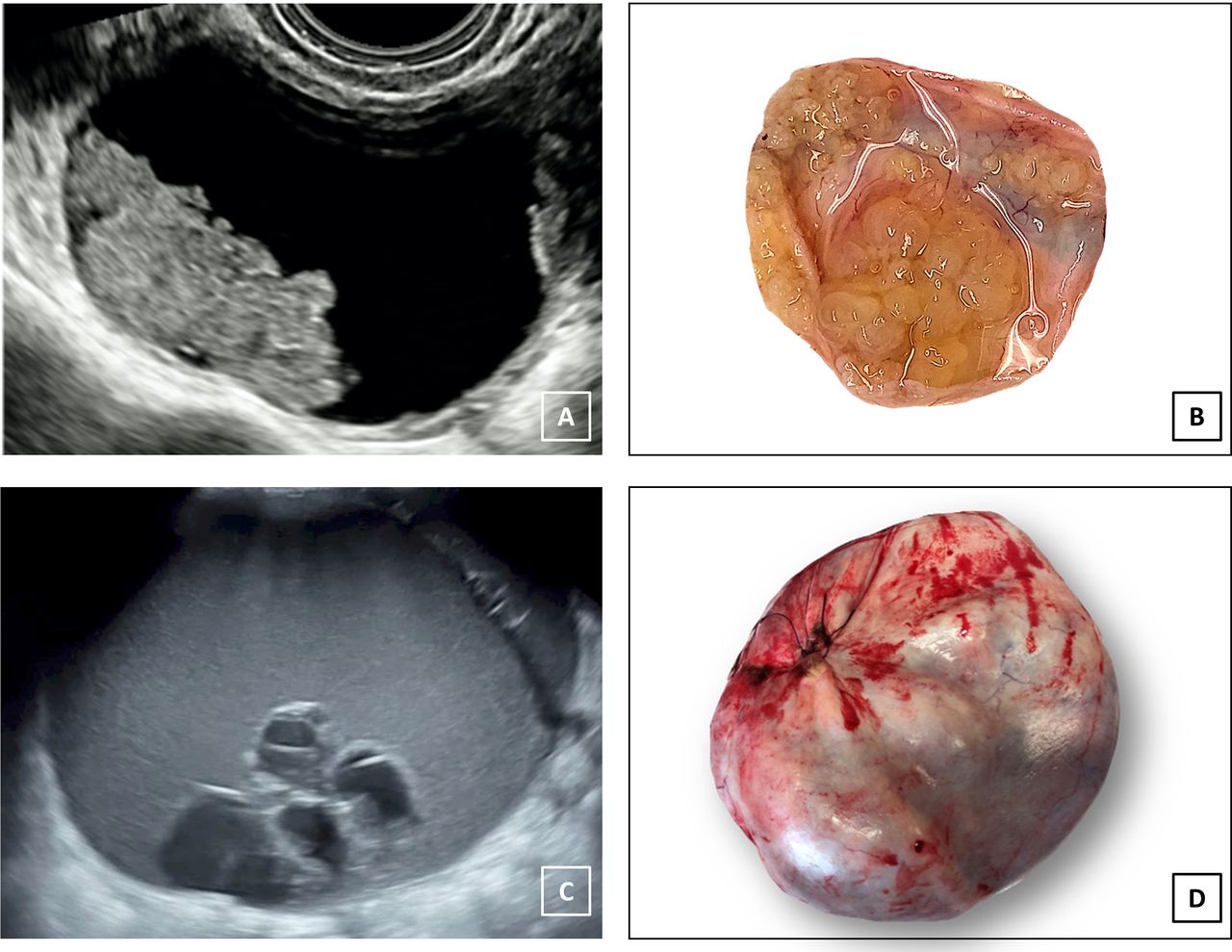
The first case is a 39-year-old patient with no family history of cancer, referred to our center for an adnexal mass incidentally detected during a routine ultrasound examination performed at another hospital. Serum levels of oncological markers were: CA125 22 U/mL (reference range: 0–35 U/mL), CA 19.9 <0.3 U/mL (reference range: 0–37 U/mL), human epididymis protein 4 (HE4) 66 pmol/L (reference range: 0–150 pmol/L), carcinoembryonic antigen (CEA) 0.42 ng/mL (reference range: 0–5 ng/mL). Transvaginal ultrasound examination showed a right unilocular solid tumor 68×39×53 mm, with anechoic content and multiple papillary projections. The largest papillary projection was 17×46 mm and it showed moderate vascularization. At ultrasound examination of the left ovary, an unilocular solid tumor 19×18×16 mm, with anechoic content and a papillary projection with moderate vascularization, was present. IOTA ADNEX model1 showed an increased risk of malignancy, and the relative risk was highest for borderline tumor (link to the IOTA ADNEX model calculator: https://www.iotagroup.org/sites/default/files/adnexmodel/IOTA-ADNEXmodel.html). Moreover, the tumor was classified as O-RADS 5.2 At laparoscopy, bilateral ovarian masses were confirmed, and a right salpingo-oophorectomy and left cystectomy were performed. The macroscopic assessment of the right mass confirmed the presence of a solid cystic tumor with multiple papillary projections. Final histology report was positive for bilateral serous borderline ovarian tumor (Figure 1).3
Ultrasound and macroscopic images of a serous borderline ovarian tumor (A, B) and of a mucinous borderline ovarian tumor (C, D).
The second case was a 57-year-old patient with no family history of cancer. During a transabdominal ultrasound examination performed for abdominal and pelvic swelling, a complex adnexal mass was detected. Serum levels of oncological markers were: CA125 22 U/mL (reference range: 0–35 U/mL), CA 19.9 <0.3 U/mL (reference range: 0–37 U/mL), CEA 0.32 ng/mL (reference range: 0–5 ng/mL). Transvaginal ultrasound examination showed a left multilocular cyst 180×110×205 mm, with more than 10 locules and low-level content. In the cyst it was possible to observe a multilocular nodule termed a ‘honeycomb nodule’.4 5 The cyst showed minimal vascularization on color Doppler examination. IOTA ADNEX model1 showed an increased risk of malignancy, and the relative risk was highest for borderline. Moreover, the tumor was classified as O-RADS 4.2 At laparoscopy, a voluminous left adnexal mass reaching the transverse umbilical plane was seen. Intra-operative frozen section of the left ovarian mass was positive for mucinous cystadenoma. Hysterectomy, bilateral salpingo-oophorectomy, and appendectomy were performed. At macroscopy, a left multilocular cystic mass with mucinous content was described.6 Final histology report was positive for mucinous borderline ovarian tumor (Figure 1) with focal intraepithelial carcinoma.7
Dott.ssa Francesca Moro is a gynecologist of Fondazione Policlinico Universitario Agostino Gemelli, IRCCS, in Rome. She is particularly involved in clinical research in ultrasound and gynecologic oncology.

Footnotes
Contributors All the authors contributed to the collection of clinical, ultrasound, macroscopic, and histological materials and in the realization of the video.
Funding The authors have not declared a specific grant for this research from any funding agency in the public, commercial or not-for-profit sectors.
Competing interests None declared.
Patient consent for publication Not required.
Provenance and peer review Commissioned; internally peer reviewed.