Article Text

Statistics from Altmetric.com
Case Presentation
An 84-year-old woman presented to an outside institution with a 1-month history of generalized malaise, anorexia, abdominal distension, shortness of breath, and unintentional 20-pound weight loss. She had a past medical history of JAK2-positive polycythemia rubra vera, stage 4 chronic kidney disease, type 2 diabetes mellitus, and hypothyroidism. Her surgical history included a total vaginal hysterectomy and bilateral salpingo-oophorectomy in 1984. Since moving to the United States in 1972, she undertook routine visits to her native country, India. The patient’s diabetes was managed with metformin/glipizide and her primary care provider had changed this regimen to sitagliptin 2 weeks prior due to worsening chronic kidney disease. On presentation, vital signs were within normal range aside from mild tachycardia. Her exam was notable for atrophy of the extremities and a protuberant abdomen with a positive fluid wave and shifting dullness. On pelvic examination, the uterus was surgically absent and there was no cul-de-sac nodularity on bimanual or rectovaginal exam.
Relevant laboratory values included: hemoglobin 11.7 g/dL, white blood cell count 9850/mm3, platelet count 468 000 mm3, blood urea nitrogen 46 mg/dL, creatinine 2.5 mg/dL, albumin 2.4 g/dL, albumin-corrected calcium 12.18 mg/dL, aspartate aminotransferase 81 U/L, alanine aminotransferase 81 U/L, carcinoembryonic antigen 1.8 ng/mL, alpha-fetoprotein 4.4 ng/mL, CA125 245 U/mL, and cancer antigen (CA) 19–9 57.4 U/mL. An initial chest radiograph provided no evidence of significant cardiopulmonary issues. A subsequent paracentesis yielded 1100 mL of fluid cytologically negative for malignancy.
Dr Johnson
What imaging is recommended given this clinical presentation, and what did it demonstrate?
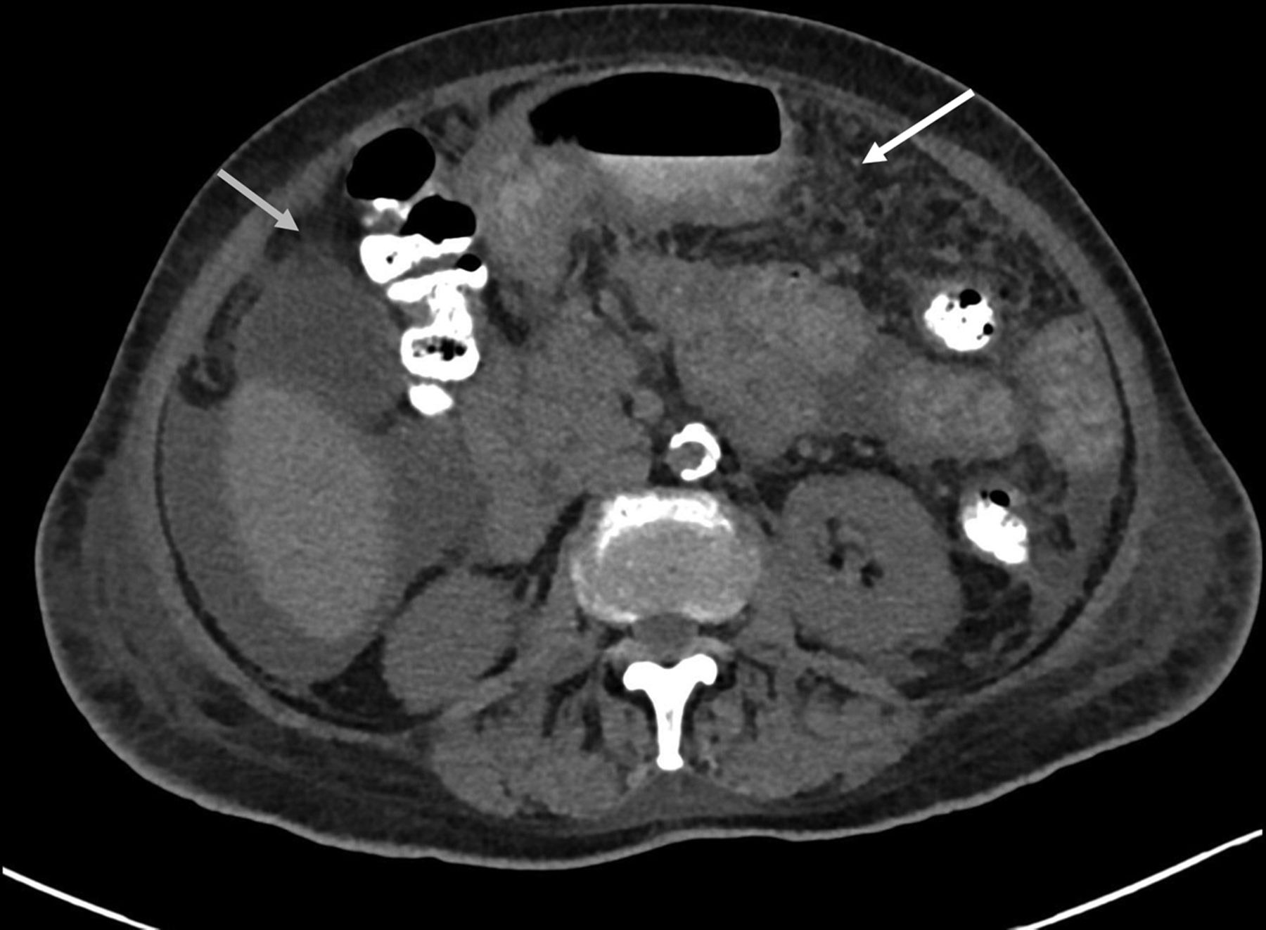
Computed tomography (CT) of the abdomen/pelvis was performed and revealed a moderate amount of ascites and an infiltrative process involving the omentum. There was no lymphadenopathy and the liver and kidneys appeared anatomically normal (Figure 1). Subsequent 18F-fluorodeoxyglucose (FDG)-positron emission tomography (PET)-CT revealed FDG-avid peritoneal thickening, nodularity throughout the peritoneal cavity, and omental fat-stranding with moderate FDG activity (standardized uptake value lean body mass (SUVlbm) max of 3.6), suggestive of peritoneal carcinomatosis (Figure 2).

CT scan of the abdomen without intravenous contrast was notable for moderate ascites (left arrow) with diffuse soft tissue stranding throughout the small bowel mesentery and greater omentum (right arrow). There was no CT evidence of an abdominopelvic mass or retroperitoneal adenopathy.

FDG PET/CT demonstrates a moderately FDG-avid area of peritoneal thickening/nodularity (white arrow). There is a moderate amount of ascites and mesenteric fat stranding mimicking a primary peritoneal carcinoma or carcinomatosis. Additional right and left internal mammary lymph nodes, as well as a right cardiophrenic lymph node, also exhibited moderate FDG-avidity in the chest.
Dr Stone
Based on this information, what would be your diagnostic approach?
In an elderly woman with a history of foreign travel, unintentional weight loss (greater than 5% of body weight over 6–12 months), ascites, hypercalcemia, and imaging concerning for carcinomatosis; malignancy (carcinomatosis or lymphomatosis) and chronic infection would be highest on the differential diagnosis. This presentation could also be seen with sclerosing peritonitis. Ovarian or peritoneal cancer are certainly high on the differential, but I am concerned about a potential infection mimicking carcinomatosis. Other diagnoses, such as end-organ damage from poorly controlled diabetes mellitus, thyroid dysfunction, congestive heart failure, hepatic dysfunction, kidney disease, and adverse medication effects should be ruled out with laboratory evaluation. Evaluation includes review of existing imaging, transthoracic echocardiogram for cardiac function, renal studies, and serologic testing for infection.
Hypercalcemia can be life-threatening, warranting urgent evaluation and management. Clinical manifestations of severe hypercalcemia include anorexia, muscle weakness, arthralgia, and renal dysfunction. Work-up entails measuring an ionized calcium, parathyroid hormone/vitamin D levels, and multiple myeloma studies.
Transthoracic echocardiogram showed normal left ventricular function with an ejection fraction of 65% and no structural abnormalities. Renal evaluation included a urine protein–creatinine ratio of 2.4, a value noted to be stable from 5 years earlier. Fourth-generation HIV testing was negative. A hepatitis panel returned positive antibodies for hepatitis A only. Evaluation of current medications revealed no conspicuous adverse side effects consistent with the patient’s clinical presentation.
The ionized calcium level was elevated at 1.7 mmol/L. Parathyroid hormone was suppressed (4 pg/mL), but parathyroid hormone-related peptide was normal (19 pg/mL). Serum and urine protein electrophoresis were negative for multiple myeloma. 25-Hydroxyvitamin D was mildly below normal range at 26 ng/mL and 1,25-hydroxyvitamin D was elevated at 91 pg/mL, suggesting low likelihood of vitamin intoxication.
Drs Fader and Salimain
What can be discerned from this clinicopathologic work-up?
Patients who present with ascites will often have a serious underlying medical problem, which can include cirrhosis complicated by portal hypertension, heart failure, nephrotic syndrome, spontaneous bacterial peritonitis, malignancy-related peritoneal carcinomatosis, or systemic granulomatous disease (sarcoidosis, peritoneal tuberculosis). In this case, the liver did not appear cirrhotic on imaging. Additionally, the patient’s unremarkable transthoracic echocardiogram was inconsistent with cardiomyopathy or valvular disease. Her non-nephrotic range proteinuria put a renal etiology lower on the differential diagnosis. An abdominal paracentesis is central to the diagnostic work-up of new-onset ascites. Peritoneal fluid can be screened for evidence of malignancy, infection, and portal hypertension. These tests may include cytology, a cell count/differential, total protein concentration, bacterial and fungal culture (infection), glucose concentration (infection and malignancy), adenosine deaminase activity (peritoneal tuberculosis), and serum-ascites albumin gradient (portal hypertension). Her initial abdominal paracentesis does not rule out malignancy, given the 20%–30% false-negative rate of peritoneal fluid sampling in the setting of ovarian cancer with malignant ascites.1Laparoscopic-guided biopsy to collect tissue for histopathologic evaluation and culture is beneficial.
The patient was transferred to our gynecologic oncology service 6 days after admission for further management of presumed primary peritoneal carcinomatosis. A diagnostic laparoscopy was performed with drainage of an additional 1500 mL of ascites. Biopsies of the omentum and diaphragm were obtained, as omental and miliary peritoneal disease was noted during surgery (Figure 3).
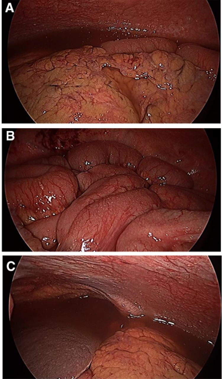
Laparoscopic images of ascites, an infiltrative process in the omentum and disseminated miliary disease as indicated by patchy thickening of the infracolic omental fat (A) and millimeter-sized white nodules studded along the small bowel serosa (B) and diaphragm peritoneum (C) .
The ascites was again devoid of malignant cells. Additional ascitic fluid results included a peritoneal fluid serum-ascites albumin gradient greater than 1.1 g/dL with a lymphocytic predominance, a white blood cell count of 7826 cells/mm3, and an absolute polymorphonuclear leucocyte count of 5488 cells/mm3 (30% of total cells). All ascitic cultures during the patient’s hospitalization were negative. The omental biopsy showed focally necrotizing, granulomatous inflammation and no histopathologic evidence of malignancy(Figure 3A). A Kinyoun stain of the same specimen showed evidence of single acid-fast bacilli (Figure 3B). An adenosine deaminase level was not obtained.

(A) Histopathologic findings of diffuse, focally necrotizing granulomatous inflammation on the omentum (black arrows). (B) Kinyoun stain with evidence of single acid fast bacilli in omental biopsy (black arrow).
The finding of acid-fast bacilli on Kinyoun staining of tissues involved with necrotizing granulomatous inflammation is highly suggestive of peritoneal tuberculosis. The patient’s recent travel to India, hypercalcemia, lymphocytic predominant ascites, and otherwise negative evaluation for alternative primary diagnoses support this. Therapy with rifampin, isoniazid, pyrazinamide, and ethambutol (RIPE) was started on post-operative day 4. An omental mycobacterial culture eventually returned positive on post-operative day 16, demonstrating pan-sensitivity to all four medications.
Dr Salimian
How can detection of mycobacteria (and differentiation from an epithelial cancer) be optimized in this setting?
Microbiologic testing can include either the Ziehl-Neelsen or Kinyoun method, as both acid-fast stains detect the mycolic acid content in the cell walls of mycobacteria with the latter method not requiring heat in the staining process.2Sensitivity of mycobacterial detection in the ascites specimen is 3% and is limited by the detection requirement of 5000 bacilli/mL. The World Health Organization (WHO)-endorsed ‘gold standard’ for mycobacterial detection is the standard culture as it requires significantly fewer organisms for detection, but it is limited by its time requirement of 4–8 weeks for final results.3
Dr Stone
What do we know about disseminated intraperitoneal tuberculosis and treatment strategies?
Ascitic fluid with a white blood cell count greater than 500 cells/mm3 with fewer than 50% polymorphonuclear leukocytes (lymphocytic predominance) supports the diagnosis of disseminated mycobacterial infection. Spontaneous bacterial peritonitis presents with a serum-ascites albumin gradient greater than 1.1 g/dL, but ascitic culture typically yields bacterial growth, and the polymorphonuclear count is usually greater than 50%. Peritoneal tuberculosis presents with signs and symptoms that mimic an advanced-stage gynecologic or gastrointestinal cancer. Fluid and tissue procurement by paracentesis and image- or laparoscopic-guided biopsy for pathology and microbiology laboratory assessment is key. Still, conventional acid-fast bacilli culture may take 4–8 weeks for identification and susceptibility testing. Postponing empiric RIPE therapy can result in further systemic dissemination and increase the risk of mortality. Hypercalcemia is present in granulomatous disease, which includes tuberculosis, and its pathology stems from an overproduction of 1,25-dihydroxyvitamin D [1,25(OH)2D3] by activated macrophages and T-lymphocytes. Treatment includes expansion of intravascular volume, corticosteroids, and hydroxychloroquine/ketoconazole for refractory cases.4
The patient was transferred to the infectious disease service to continue on daily RIPE therapy and vitamin B6, per Centers of Disease Control guidelines. Her ionized calcium levels peaked at 1.93 mmol/L, and therapeutic interventions included fluids, furosemide, pamidronate, calcitonin, methylprednisolone, and hydroxychloroquine. She had a prolonged inpatient course with altered mental status, electrolyte abnormalities, uremia, and metabolic acidosis. During her fourth week of RIPE treatment, her condition acutely deteriorated. She became increasingly altered and developed shock. Her care team initiated hemodynamic support and broadened her antibiotic coverage. Plasma lactate was 13 mmol/L on basic laboratory testing. A non-contrast head CT was negative and a CT of the chest, abdomen, and pelvis showed marked anasarca, increasing ascites, and new bilateral pleural effusions. There was no evidence of bowel obstruction or perforation. Despite initial resuscitation, shock rapidly evolved to fatal multi-organ failure and she succumbed to suspected intestinal ischemia.
Drs Fader and Azadi
With an elevated CA125 and imaging findings consistent with carcinomatosis, why can one not presume that this always represents malignancy?
This is a complex clinical scenario as peritoneal tuberculosis mimicked ovarian/peritoneal carcinoma in several ways in this case. Both entities can demonstrate multiple nodules, thickening of tissues, plaque-like disease, and ascites on imaging as well as an elevation of CA125 on serum testing. CA125 is a coelomic epithelial glycoprotein that is a non-specific marker of peritoneal inflammation in processes such as ovarian cancer, endometriosis, pelvic inflammatory disease, pancreatitis, and cirrhosis. It is elevated in tuberculous peritonitis, and its downtrend with therapy can be used to monitor response to treatment.5 6
Symptoms of both peritoneal cancer and intraperitoneal infection, such as tuberculosis, are similar and confounding. They include fatigue, pain, weight loss, anorexia, fevers, abdominal distension, and diarrhea/constipation. Physical exam can include palpation of a pelvic mass or abdominal nodularity. As gynecologic oncologists we must have a reasonable index of suspicion if multimodal evaluation provides more questions than answers. A distinguishing note in this case was the lack of evidence regarding malignancy in the ascitic specimen. The elevated CA125 and carcinomatosis on imaging were red herrings that misled the initial primary care team.
Detection of Mycobacterium tuberculosis includes ascitic fluid adenosine deaminase and gamma-interferon. Levels greater than 30 U/L and 3.2 U/mL provide a sensitivity/specificity of 93%/96% and 93%/98%, respectively.7Both markers are indicative of T-lymphocyte activity caused by mycobacterial stimulation. Although an ascitic adenosine deaminase level was not obtained, the surgically obtained tissue biopsy was warranted and should always be performed in this setting prior to therapy initiation for a systemic disease.
In patients with peritoneal tuberculosis, an abnormal chest radiograph is found in 21%–83% of cases, with active pulmonary tuberculosis present in about 14% of cases.3The most common finding on CT scan is high-density ascites with evidence of floating debris and fibrin stranding in the exudative material. CT scan may also disclose lymphadenopathy and omental caking.8As demonstrated by this case, the imaging findings are indistinguishable from epithelial ovarian carcinomatosis, primary peritoneal cancer, peritoneal sarcoidosis, and lymphomatosis.
Dr Stone
Closing Summary
This case highlights the fact that ascites, hypercalcemia, and a recent travel history to areas of the world endemic to tuberculosis are early clues that mycobacterial infection may be masquerading as peritoneal carcinomatosis. Gynecologic oncologists should consider this in their differential diagnoses. Pre-operative imaging was consistent with carcinomatosis, but in the setting of ascitic cytology negative for malignancy, diagnostic laparoscopy with tissue biopsies are paramount for the correct diagnosis. Intra-operative findings can be categorized as such: (1) wet type with characteristic ascites, miliary nodules, and hyperemic peritoneum as seen in our case (Figure 4); (2) dry type with ‘plastic-like’ findings and adhesions; and (3) fibro-adhesive type with caseous collections and adhesions.9Intestinal obstruction occurs in fewer than 10% of cases. Bowel perforation occurs in 1%–15% of cases, and most commonly occurs in the terminal ileum due to high-density lymphoid aggregates known as Peyer’s patches.9–11
An ascitic adenosine deaminase level was not obtained in this case and could have facilitated an earlier diagnosis. Despite our patient’s advanced age and mildly elevated transaminase levels on presentation, the benefits of prompt administration of empiric tuberculosis therapy outweighed the risks, as it optimizes the chance of cure and minimizes the risk of relapse. Risks of delaying therapy can be catastrophic due to evolving toxicity to the gastrointestinal tract from untreated disease culminating in multifocal obstruction, perforated viscus, and ischemia. Initiation of anti-tuberculosis therapy is not without risk either, as intestinal obstruction can worsen due to healing by cicatrization.9Given that peritoneal tuberculosis can mimic many diverse clinical conditions, including epithelial ovarian or peritoneal cancers, misdiagnosis and treatment delays are common jeopardies. Considering the time period was 13 days from initial presentation to start of RIPE therapy, there remains the unanswered question of when pharmacotherapy would have changed the patient’s clinical outcome. An evidence-based approach to answering this question is not feasible. Instead, patient travel history, laboratory and radiological findings, ascitic studies, and intraperitoneal biopsies all play an important role in correctly identifying and treating this life-threatening infection.
Footnotes
Twitter @amandanfader, @MaryAnnBWilbur
Contributors All authors have contributed equally including conceptualization, data curation, validation, and writing/editing the original draft.
Funding The authors have not declared a specific grant for this research from any funding agency in the public, commercial or not-for-profit sectors.
Competing interests None declared.
Patient consent for publication Not required.
Provenance and peer review Commissioned; internally peer reviewed.