Article Text

Download PDFPDF

2022-RA-959-ESGO Pelvic sentinel lymph nodedistribution; the final outcome of the Sentix trial (CEEGOG-CX01; ENGOT-CX2; NCT02494063)
Free
  1. Roman Kocian1,
  2. Christhardt Koehler2,
  3. Sylva Bajsova3,
  4. Klara Benesova4,
  5. Ignacio Zapardiel5,
  6. Giampaolo Di Martino6,
  7. Luc van Lonkhuijzen7,
  8. Borek Sehnal8,
  9. Octavio Arencibia Sanchez9,
  10. Blanca Gil Ibanez10,
  11. Fabio Martinelli11,
  12. Jiri Presl12,
  13. Lubos Minar13,
  14. Radim Marek14,
  15. Peter Kascak15,
  16. Pavel Havelka16,
  17. Martin Michal17,
  18. Toon van Gorp18,
  19. Kristyna Nemejcova19 and
  20. David Cibula1
  1. 1Gynecologic Oncology Center, Department of Obstetrics and Gynecology, First Faculty of Medicine, Charles University and General University Hospital in Prague, CEEGOG, Prague, Czech Republic
  2. 2Department of Special Operative and Oncologic Gynaecology, Asklepios-Clinic Hamburg, Hamburg, Germany
  3. 3Department of Obstetrics and Gynecology, University Hospital Ostrava, CEEGOG, Ostrava, Czech Republic
  4. 4Institute of Biostatistics and Analyses, Faculty of Medicine, Masaryk University, Brno, Czech Republic
  5. 5Department of Obstetrics and Gynecology, La Paz University Hospital, Madrid, Spain
  6. 6Department of Obstetrics and Gynecology, Unit of Gynecologic Oncology Surgery, San Gerardo Hospital, Monza, Italy
  7. 7Centre for Gynecologic Oncology, Academic Medical Centre, Amsterdam, Netherlands
  8. 8Department of Obstetrics and Gynecology, University Hospital Bulovka, First Faculty of Medicine, Charles University, CEEGOG, Prague, Czech Republic
  9. 9Department of Gynecologic Oncology, University Hospital of the Canary Islands, Las Palmas de Gran Canaria, Spain
  10. 10Unit of Gynecological Oncology, Institute Clinic of Gynecology, Obstetrics and Neonatology (ICGON), Hospital Clinic of Barcelona, Barcelona, Spain
  11. 11IRCCS Foundation National Cancer Institute in Milan, Milan, Italy
  12. 12Department of Gynaecology and Obstetrics, University Hospital Pilsen, Charles University, CEEGOG, Pilsen, Czech Republic
  13. 13Department of Gynecology and Obstetrics, Faculty of Medicine, Masaryk University, CEEGOG, Brno, Czech Republic
  14. 14Department of Obstetrics and Gynecology, Faculty of Medicine and Dentistry, Palacky University, University Hospital Olomouc, CEEGOG, Olomouc, Czech Republic
  15. 15Department of Obstetrics and Gynecology, Faculty Hospital Trencin, CEEGOG, Trencin, Slovakia
  16. 16Department of Obstetrics and Gynecology, KNTB a.s, CEEGOG, Zlin, Czech Republic
  17. 17Department of Obstetrics and Gynaecology, Hospital Ceske Budejovice, JSC, CEEGOG, Ceske Budejovice, Czech Republic
  18. 18Department of Gynecology and Obstetrics, University Hospital Leuven, Leuven Cancer Institute, BGOG, Leuven, Belgium
  19. 19Institute of Pathology, First Faculty of Medicine, Charles University and General University Hospital, Prague, Czech Republic

Abstract

Introduction/Background Over the last twenty years, data from more than 2000 patients from thirty studies on sentinel lymph node (SLN) mapping in early-stage cervical cancer were published. Many of these reports come from small single-centre studies or retrospective data from the time when detection rates were much lower. We present final results on SLN mapping from the Sentix study, the largest prospective cohort study of more than 700 patients.

Methodology Eligible were patients with cervical cancer stages T1a1 L1 – T1b2 (<4 or ≤2 cm for fertility sparing), common tumour types and no suspicious lymph nodes on preoperative imaging. All detection techniques (blue dye, radiocolloid, indocyanine green) and combinations were allowed. Preoperative lymphoscintigraphy was not required and not used. All approaches, laparotomy, laparoscopy, or robotic surgery were acceptable. Intraoperatively pelvic (external iliac, interiliac, common iliac, presacral) and low paraaortic regions were examined for the presence of SLN. All patients with successful bilateral SLN detection and a completed postoperative data continued in the study.

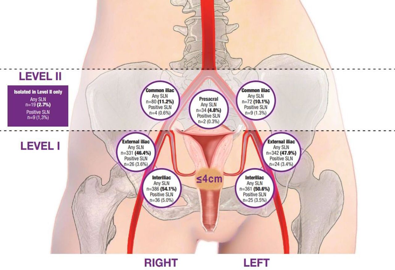
Results Final cohort of 714 patients were analysed, enrolled between 2016–2020 in 47 centres and 18 participating countries. Bilateral SLN detection rate reached 92.3% with the median of 3 SLNs per patient. All SLNs were detected in the pelvis, no SLN in the low paraaortic region. The majority (97.3%) were localized in the pelvic level I, below the interiliac bifurcation. There was an extremely low rate (1.3%) of isolated positive SLNs in pelvic level II. No laterally distinct distribution of SLNs was found.

Abstract 2022-RA-959-ESGO Figure 1

Conclusion During SLN biopsy, surgical pelvic dissection should focus on the bilateral anatomical area below the interilical bifurcation, the external iliac vessels region, and the obturator fossa, where SLNs are most frequently located. Occurrences outside this region are rare with an extremely low risk of isolated metastatic SLN in the pelvic level II.

Statistics from Altmetric.com

Request Permissions

If you wish to reuse any or all of this article please use the link below which will take you to the Copyright Clearance Center’s RightsLink service. You will be able to get a quick price and instant permission to reuse the content in many different ways.