Article Text

- http://orcid.org/0000-0003-3126-9400Andrea Rosati1,
Dr. Andrea Rosati is a gynecologist oncologist of Fondazione Policlinico Universitario Agostino Gemelli, IRCCS, in Rome. He is particularly involved in clinical research and surgical anatomy. He is currently a fellow in the 2nd Level International Master in Gynecologic Oncology accredited by ESGO society.

- Agostino Maria De Rose2,
- Evis Sala3,
- Felice Giuliante2,4,
- http://orcid.org/0000-0003-2758-1063Giovanni Scambia1,4 and
- http://orcid.org/0000-0001-5579-335XAnna Fagotti1,4
- 1 Department of Woman and Child Health and Public Health, Fondazione Policlinico Universitario A. Gemelli, IRCCS, Rome, Italy
- 2 Hepatobiliary Surgery Unit, Fondazione Policlinico Universitario A. Gemelli, IRCCS, Rome, Italy
- 3 Department of Radiology, Cambridge University, Cambridge, Cambridgeshire, UK
- 4 Università Cattolica del Sacro Cuore Facoltà di Medicina e Chirurgia, Rome, Italy
- Correspondence to Professor Giovanni Scambia, Fondazione Policlinico Universitario Agostino Gemelli IRCCS, Rome 00168, Italy; giovanni.scambia{at}policlinicogemelli.it
Statistics from Altmetric.com
Summary
The combination of emerging targeted therapies and technological advancement in surgical procedures supports the trend towards a prolonged survival in advanced ovarian cancer patients. Tumor localization in the liver area has become more frequent and more challenging to manage during the natural course of the disease.1 2
We developed an anatomo-surgical classification for ovarian cancer metastases in the liver area3 aiming to provide a standardized nomenclature during pre-operative plan and surgical report.
Disclaimer: this video summarises a scientific article published by BMJ Publishing Group Limited (BMJ). The content of this video has not been peer-reviewed and does not constitute medical advice. Any opinions expressed are solely those of the contributors. Viewers should be aware that professionals in the field may have different opinions. BMJ does not endorse any opinions expressed or recommendations discussed. Viewers should not use the content of the video as the basis for any medical treatment. BMJ disclaims all liability and responsibility arising from any reliance placed on the content.
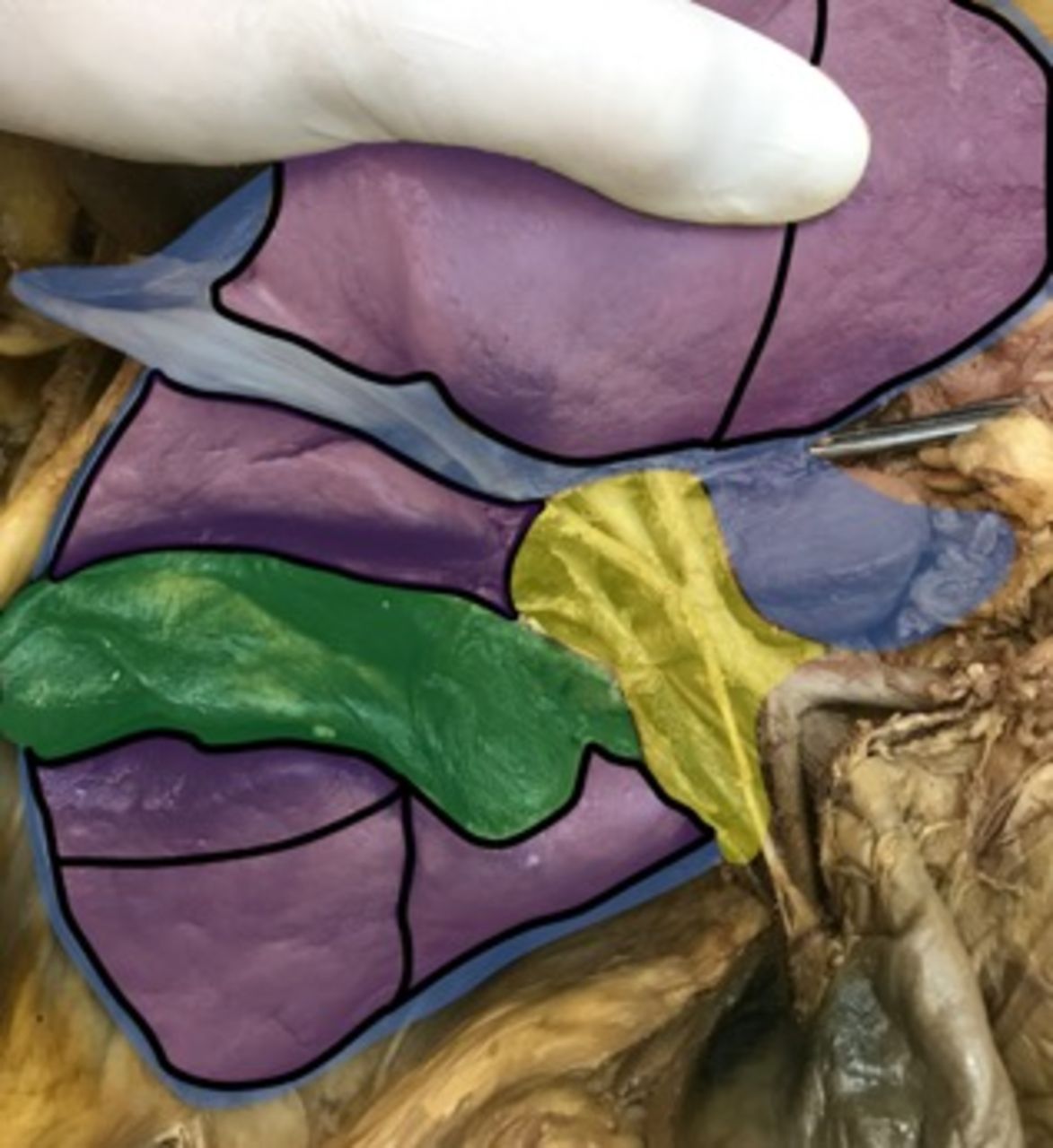
We identified five conceptually distinct anatomical areas using the three-dimensional anatomical model4 and surgical videos (Figure 1).
Anatomo-surgical classification of ovarian cancer metastases in the liver area. Purple area: type 1 or Glisson’s capsule. Blue area: type 2 or ligamentous. Green area: type 3 or gallbladder. Yellow area: type 4 or hepatic hilum. Black lines: type 5 or parenchymal (subdivided by liver segment).
Our anatomo-surgical classification consists of five distinct categories:
Type 1 or Glisson’s capsule: superficial metastases involving the Glisson’s sheet with no parenchymal infiltration.
Type 2 or ligamentous: this is a heterogeneous group including carcinomatosis along the lines of reflection between the liver and surrounding organs (falciform ligament, round ligament, coronary and triangular ligament, Arantii and hepato-gastric ligament).
Type 3 or gallbladder: neoplastic nodules located along the gallbladder surface and fossa.
Type 4 or hepatic hilum: porta hepatis is considered as a single entity due to the potential neoplastic involvement from the peritoneal site (hepato-duodenal ligament and Rouviere’s sulcus) and lymphatic site (portal triad lymph nodes).
Type 5 or parenchymal: divided into ‘superficial’, infiltrating <1 cm in depth, and ‘intra-parenchymal’, traditionally classified according to the liver segment.
Our classification represents a useful guide while planning the surgical strategy for advanced ovarian cancer metastases in the liver area, assigning the specific procedure within a multidisciplinary team, based on surgical competence. Especially in types 3, 4, and 5 metastases a requirement for hepatobiliary surgeon should be anticipated.
The standardization of nomenclature allows an easy exchange of surgical information for education and scientific purposes, which are otherwise difficult to interpret and compare.
The identification of specific risks and strategies related to each anatomical localization provides a didactic and effective tool.
Ethics statements
Patient consent for publication
Acknowledgments
We acknowledge Doctors: Francesco Fanfani, Barbara Costantini, Angelica Naldini, Valerio Gallotta, Lucia Tortorella, Luigi Turco, Giorgia Monterossi, Giovanni Ardito, Claudio Lodoli, Francesco Santullo, Carlo Abatini and Fabio Pacelli for their continuous surgical and educational effort and for their great dedication to patients. We acknowledge Arianna Catena for her contribution in the voice-over of the video.
Dr. Andrea Rosati is a gynecologist oncologist of Fondazione Policlinico Universitario Agostino Gemelli, IRCCS, in Rome. He is particularly involved in clinical research and surgical anatomy. He is currently a fellow in the 2nd Level International Master in Gynecologic Oncology accredited by ESGO society.
Footnotes
Twitter @annafagottimd
Contributors Conceptualization: AF, AR; Supervision: AF, GS, ES; Writing - review & video editing: AR, AF, AMDR; Review & editing: AF, GS, FG, ES, AR, AMDR; All authors read and approved the final manuscript.
Funding The authors have not declared a specific grant for this research from any funding agency in the public, commercial or not-for-profit sectors.
Competing interests None declared.
Provenance and peer review Commissioned; externally peer reviewed.