Article Text

Abstract
Introduction/Background*Sarcomas represent 1% of gynecological tumors and between 3-7% of uterine neoplasms. Given its low incidence, the available evidence and literature is limited. We provide our data as a self-assessment and analysis of our healthcare practice
Methodology Retrospective study of patients with uterine sarcomas diagnosed and treated in CHUIMI in the period 2009-2018. We included epidemiological variables, stage at diagnosis, treatment, anatomo-pathological features, follow-up and current status of the patients.
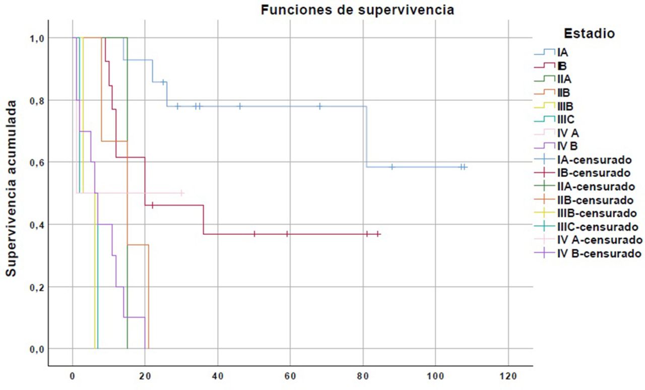
Result(s)*The total number of patients diagnosed with uterine sarcoma was 47, with a mean age of 56.8 years [31-85]. 42.6% of patients were in an advanced stage at diagnosis [Stage I 57.4% (27), II 8.5% (4), III 8.6% (4) and IV 25.6% (12)].
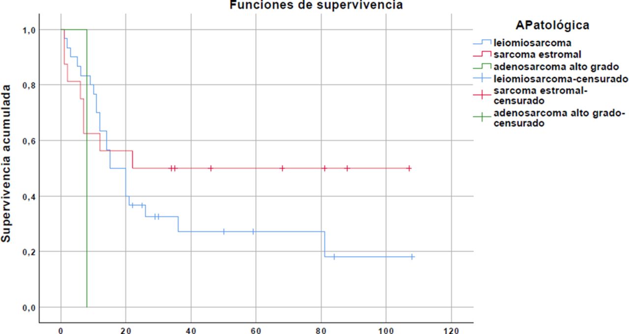
Regarding histology, we found that 63.8% (30) were Leiomyosarcomas, 34% (16) were Stromal sarcomas and 2.1% (1) High-grade adenosarcomas. Overall survival at 5 years is 36.17% with a median of 20 months. After 5 years of follow-up, 27.2% of leiomyosarcomas lived (median 15 months), 50% of sarcomas stromal (median 22 months), and none of the high-grade adenosarcomas (median 8 months). Globally, in relation to the stage of the disease at diagnosis, after 3 years of follow-up 59.25% of the stages I survived (stable up to 5 years), and none of stages II, III or IV survived.
Regarding the type of treatment, 87.2% of the patients underwent surgery (61.7% LPT; 23.4% LPC). Of these, only 21.3% did not receive adjuvant treatment (34% RT, 17% QT, 14.9% RT + QT). Globally, 21.3% of the patients relapse (most frequently in the lung, 8.5%, followed by local recurrence 6.4%, abdominal 4.3% and bone 2.1%) compared to 51.1% who progress.
There was fragmentation of the surgical piece in 19.1% (no morcellation). 34% of tumors are> 10 cm. 31.9% had a low mitotic index (<5). 29.8% presented lymphovascular invasion
Conclusion*Our epidemiological and survival data coincide with what has been published in the literature. It is important to provide evidence on a pathology that, although rare, presents so much impact on our patients, thus contributing to achieve better clinical practices.