Article Text

Abstract
The major tenets in accurately assessing tumor size in patients with early stage cervical cancer currently include physical examination, imaging studies, and pathologic evaluation. It is estimated that when comparing clinical stage based on physical examination and final pathology, the concordance diminishes as stage increases: 85.4%, 77.4%, 35.3%, and 20.5% for stage IB1, IB2, IIA, and IIB, respectively. Vaginal involvement and larger tumor diameter are considered the main causes of stage inaccuracy. When considering imaging studies, magnetic resonance imaging (MRI) provides the highest level of accuracy in the assessment of cervical tumor size. Its accuracy in determining tumor location within the cervix is approximately 91% and in predicting tumor size 93%. MRI imaging is also significantly more accurate in measuring tumor size, delineating cervical tumor boundaries, and local tumor extension when compared with computed tomography (CT) scan. When comparing with pelvic ultrasound, the accuracy of both imaging techniques (MRI and pelvic ultrasound) in the assessment of tumor size in small versus large tumors is comparable. Pertaining to pathology, the depth of invasion should be measured by convention from the nearest surface epithelium, which equates to tumor thickness. In the setting where tumor is found both in the conization and hysterectomy specimen, the horizontal extent should be measured by summing the maximum horizontal measurement in the different specimens and the depth of invasion measured as the maximum depth in either specimen. A new pattern-based classification for endocervical adenocarcinomas recommends the description of patterns of invasion for human papillomavirus (HPV)-related adenocarcinomas as this is associated with differing risks of lymph node involvement.
- cervical cancer
- cervix uteri
- uterine cervical neoplasms
Statistics from Altmetric.com
Introduction
Accurate staging of patients with cervical cancer is crucial for appropriate treatment planning. The first reports on staging of cervical cancer date back to 1928.1
The currently recognized and established staging system was implemented as the FIGO (International Federation of Gynecology and Obstetrics) staging system in 1958. Tumor size has consistently been a key component in the stage of disease in patients diagnosed with cervical cancer. Over time, there have been numerable reiterations of the staging classification, particularly for “early-stage disease”. In 1994, the staging system drew its attention to the differentiation between different tumor sizes. In previous FIGO staging reviews, stage I included all tumors confined to the cervix (FIGO 1950 and 1971), later subdividing stage I into IA (preclinical invasive carcinoma) and IB (FIGO 1974). In 1985, FIGO provided further modification and divided stage IA into IA1 (minimal microscopically evident stromal invasion) and IA2 (lesions detected microscopically that can be measured, depth of invasion ≤5 mm and horizontal spread ≤7 mm). This sub-staging was based on the fact that there was increasing evidence that tumor size was associated with prognosis. Then in 1994, further clarification was provided by FIGO to expand on the definition of this subcategory, stage IA1 (stromal invasion ≤3 mm and horizontal spread ≤7 mm) and stage IA2 (stromal invasion >3 mm to ≤5 mm and horizontal spread ≤7 mm). Specifically, “bulky” stage IB was considered an important prognostic factor in stage I disease with a 90% survival for patients with stage IB in comparison to 50–60% for IB “bulky” tumors. Therefore, stage IB was divided into IB1 (≤4 cm) and IB2 (>4 cm).2 This change also integrated measurement of tumor diameter in three dimensions: anteroposterior, lateral, and craniocaudal. These measurements were to be obtained by bimanual examination through a rectovaginal examination under anesthesia. More recently, almost 60 years after the original FIGO staging system was established, there continues to be an emphasis on the relevance of tumor size in cervical cancer. The 2018 FIGO classification has subdivided stage IB further into stage IB1 (<2 cm), IB2 (≥2–4 cm), and IB3 (≥4 cm).3 Another noteworthy change in the 2018 classification is the fact that prior to this change, staging of cervical cancer relied solely on pelvic examination and limited imaging studies; however, the most recent classification allows, when available, for imaging and pathology to help designate the stage of the patient.
Tumor size is a major determinant when deciding on treatment recommendations, and in patients with early-stage disease it dictates whether a patient is a candidate for conservative surgery, radical hysterectomy, or chemotherapy and radiation.4 The accurate assessment of tumor size, as well as other tumor characteristics, such as parametrial invasion, depth of invasion, and lymph node metastases, are key factors in determining the need for adjuvant therapy. Tumor size also provides a prognostic value, as large tumors have long been known to predict risk for distant spread of disease and poor prognosis in cervical cancer.5 The 5 year survival rate declines from 91.6% (95% CI 90.4% to 92.6%) for patients with tumors ≤2 cm, 83.3% (95% CI 81.8% to 84.8%) for tumors >2 to ≤4 cm, and 76.1% (95% CI 74.3% to 77.8%) for >4 cm tumors.6 Studies have found that primary tumor size >2 cm predicts parametrial invasion (p=0.001)7 with 13% parametrial involvement in patients with tumors >2 cm and 1.3% in those with tumors <2 cm.8
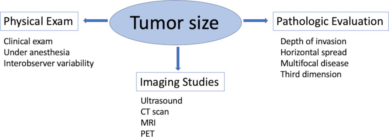
Although there is strong emphasis on the relevance and importance of accurate determination of tumor size in patients with early cervical cancer, there are still a number of discrepancies, unanswered questions, and gaps when determining criteria for tumor size measurements. One would consider that given the emphasis placed on this variable when determining discussions on prognosis, type of adjuvant therapy, and stage of disease, there would be a standardized system based on concrete and objective criteria that would allow for uniform and consistent determination of tumor size designation. Today, there are significant gaps and disagreements among methods and strategies used by gynecologic oncologists and pathologists pertaining to the clinical measurement of the tumor, there are disparities in the measurement depending on the type of preoperative imaging, and there is a lack of consensus regarding tumor size in patients who have undergone conization prior to surgery who have residual disease on hysterectomy specimen. The aim of this review is to evaluate the current literature on tumor size assessment in patients with early-stage cervical cancer as measured by pelvic examination, preoperative imaging, and pathology (Figure 1).
Tumor size assessment in early-stage cervical cancer. CT, computed tomography; MRI, magnetic resonance imaging, PET, positron emission tomography.
Physical Examination
The physical examination remains a cornerstone of cervical cancer staging; however, determination of tumor size by physical examination is fraught with a number of challenges and it is certainly prone to inaccuracy, as pelvic examination largely depends on the examiner’s experience and ability to perform a complete and thorough exam.9 A number of factors might impact this assessment: among these include, but are not limited to, body habitus, patient anxiety and discomfort, and symptoms that may preclude an adequate evaluation.10 The location of tumor is also important in the accuracy of assessing tumor size by pelvic examination, given that for tumors primarily in the endocervix, it may be difficult to determine tumor size from a pelvic examination, as the craniocaudal extension may not be easy to estimate with palpation. Also, bulky tumors or ulcerative tumors are difficult to evaluate. In addition, the clinical assessment of tumor size is based on a subjective assessment, rather than an exact measure. Tumor size is often described as a range, such as 2–3 cm. Such assessment may not be as impactful for small tumors (1–2 cm); however, when dealing with larger tumors, in the proximity of 4 cm, such discrepancies may impact whether a patient undergoes surgery versus chemotherapy and radiation. One of the first descriptions on tumor size determined by clinical examination was Van Nagell et al11 in 1971, who proposed that physical examination was generally associated with an underestimation of staging. After comparing clinical staging with final pathology in 125 patients with cervical cancer, only 83 (66%) patients were staged correctly. The investigators noted that clinical staging accuracy was highest in patients with stage I disease (78%) and lowest in patients with stage III (25%) disease. Understaging was primarily because of extension of tumor into the bladder or rectum. The authors emphasized that pelvic structures relevant to the determination of cervical tumor size were generally inaccessible, even when patients were fully anesthetized. The authors concluded that “clinical staging is at best an inaccurate estimation of the extent of cervical carcinoma”. In 1975, Averette et al12 proposed that, compared with surgical staging, clinical FIGO staging was subject to errors as high as 26% for stage IB, 45% for stage IIA, 55% for stage IIB, 66% for stage IIIA, and 94% for stage IIIB.
Mayr et al13 performed a prospective study including 43 patients with advanced cervical cancer. One of the objectives of the study was to correlate tumor measurements by physical examination versus magnetic resonance imaging (MRI). The study found that tumor size (maximum diameter, average diameter, and volume) by clinical exam and MRI did not correlate well (r=0.51, 0.61, and 0.58, respectively), with the maximum diameter being the most inaccurate measurement (r=0.51). Tumors were classified as small (<40 cm3), intermediate (40–99 cm3), and large (≥100 cm3) correlating to tumors <4 cm, 4–6 cm, and >6 cm, respectively). The correlation of tumor size was highest in small tumors, 71%, and decreased to 54% for intermediate, and 29% for large tumors. In general, physical examination tends to overestimate tumors measuring 4–6 cm. Hoffman et al14 performed an observational study to determine whether pelvic examination identified factors that suggested the need for adjuvant radiotherapy. Pathologic findings were compared with pelvic examination. Most examiners had 11 to 15 years of experience. The authors found that the accuracy of the clinical examination was approximately 50% for tumor diameter (±25%). Although the study found that the likelihood for adjuvant treatment had a significant association with the number of risk variables identified at examination, the authors concluded that for stage IB-IIA cervical cancer, clinicians who make treatment decisions based on examination findings should understand the limitations of this evaluation.
Another potential issue when relying on physical examination when staging patients with cervical cancer is the difference in assessing tumor size between surgeons, thus highlighting the impact of interobserver variability. One of the first publications on this topic dates back to 1953 and showed that staging may vary as much as 10% among examiners.15 In 2009, Qin et al16 performed a retrospective analysis of 818 patients with stage IB-IIB (FIGO 1994) cervical cancer treated with primary surgery. The authors evaluated the discrepancy between clinical stage and pathological results. The pelvic examination was performed without anesthesia by two certified gynecologic oncologists. The pTNM stage (pathological TNM classification) was used to determine the pathological stage and all patients were assigned a pT category. The overall concordance between clinical stage and pT was 53% (434 of 818), with a kappa (κ) value of 0.409 (p<0.001). The overestimation of the FIGO stage was 37% (305 of 818 patients) and the underestimation was 10% (79 of 818 patients). The concordance diminished as stage increased: 85.4%, 77.4%, 35.3%, and 20.5% for stage IB1, IB2, IIA, and IIB, respectively. The authors concluded that several discrepancies exist between clinical stage and pathological results and that for patients with cervical cancer, clinical stage alone is not reliable. Tummers et al17 evaluated 86 patients who underwent examination under anesthesia before primary treatment by at least three gynecologic oncologists, and evaluated the level of agreement in terms of tumor size, vaginal involvement, parametrium and rectal involvement, FIGO stage, and proposed therapy. For tumor size, the level of agreement was moderate (κ=0.42, p=0.07; 0.41–0.60 was considered as moderate agreement). The authors also found that for patients who underwent surgery, 33% were understaged by clinical-based evaluation.
Canaz et al5 retrospectively reviewed 129 patients with FIGO 2009 stage IB1-IIA2 who underwent a radical hysterectomy. When evaluating tumor size, they found that the mean maximal tumor diameter measured in the pathology specimen (30 mm, range 8–75 mm) was larger than the clinically estimated maximal tumor diameter (28 mm range, 8–55 mm) (p=0.016). The authors highlighted that endophytic growth is one of the most important preoperative features reflecting deep infiltration and parametrial compromise, which might be unrecognized in the pelvic examination.
A recently published large multicenter retrospective study including 12 681 patients from China showed that the overall accuracy of clinical staging was 70%, with 21% of patients clinically over-staged and 9% clinically under-staged. The accuracy of clinical staging worsened with higher clinical stage, 87.5% for FIGO 2009 stage IB1 and 26% for stage IIA2. Vaginal involvement and larger tumor diameter were the main causes of stage inaccuracy (62% and 25%, respectively). In a sub-analysis of clinical understaging, maximal tumor diameter was the primary cause of tumor understaging (47.5%). The authors concluded that the quality of staging cervical cancer was suboptimal before the integration of advanced imaging techniques as currently proposed by the 2018 FIGO staging system.18
The evidence thus suggests that clinical examination, although a standard and necessary step in the initial evaluation of patients with cervical cancer, is limited in its ability to reliably ascertain tumor size with accuracy. It is imperative that in addition to the pelvic examination, tools such as imaging studies are used to complement the information gathered in designating stage and implementing management strategy (Table 1).
Accuracy of physical examination and imaging methods in measuring tumor size
Imaging Studies
Prior to the 2018 FIGO staging update, besides physical examination, the only additional evaluation tools approved by previous FIGO staging classifications for cervical cancer were colposcopy, radiologic studies such as chest radiography, intravenous urography, and barium enema, and endoscopic studies such as cystoscopy and/or sigmoidoscopy.19 These diagnostic tests were evaluated by a prospective study performed by the American College of Radiology Imaging Network/Gynecologic Oncology Group including 25 sites in the USA from 2000 to 2002. A total of 197 early stage (FIGO 1994 IB) cervical cancer patients scheduled for surgery were included. The study showed a decreased in the use of cystoscopy (8.1%) and sigmoidoscopy or proctoscopy (8.6%) which was significantly lower than in 1988 to 1989 (p<0.0001). Intravenous urography was used in only 1% of patients in comparison to 42% in 1988 to 1989 and 91% in 1983. No patients included in the analysis had a barium enema or lymphangiography. Only 26.9% of patients had examination under anesthesia for clinical staging. The study concluded that there was a large discrepancy between the diagnostic test recommended by FIGO and tests actually used for cervical cancer staging, thus suggesting a need to re-evaluate recommendations.20
The most recent FIGO 2018 classification allows for the incorporation of imaging studies such as ultrasound, computed tomography (CT), MRI, positron emission tomography (PET), PET-CT, and MRI-PET in the staging of patients with cervical cancer.3 However, one must recognize that there is significant variation in the capacity to accurately assess the extent of disease when comparing different imaging modalities.
MRI
MRI has long been considered the method of choice in the primary evaluation of patients with cervical cancers.21 One of the first studies evaluating the role of MRI in determining cervical cancer staging showed that the accuracy of clinical staging for early stage tumors was 79% compared with 81% for MRI, but when patients with early stage disease (IB1-IIA1) were excluded, the accuracy of clinical staging decreased to 53%. The accuracy in determining tumor location within the cervix was 91% and in predicting tumor size was 93%.22 MRI has high accuracy in determining tumor size with <5 mm discrepancy between the largest diameter measured on MRI versus pathologic evaluation in 70–90% of patients.23 24 A potential pitfall in the evaluation of tumor size with MRI imaging is the differentiation between peritumoral edema and actual tumor. The inclusion of diffusion-weighted imaging in pelvic MRI protocols has helped overcome this barrier.25 In addition, it may enable detection of tumors smaller than 1 cm.26–28 MRI is also significantly more accurate in measuring tumor size, delineating cervical tumor boundaries, and local tumor extension when compared with CT scan because of its distinctive tissue contrast and multiplanar capability, in contrast to CT which is limited by its poor tissue contrast.29
CT Scan
CT is a widely available imaging modality, with short scanning time, lack of bowel motion artifacts, few contraindications, and high spatial resolution images. However, it does have unsatisfactory soft-tissue contrast resolution even with the latest technology and intravenous contrast. Cervical tumors are generally indistinguishable from adjacent structures (isodense). Even large tumors may only appear as a non-specific cervical enlargement. With a reported overall staging accuracy of 53%,30 CT scan is not considered the ideal imaging technique for the detection of cervical carcinomas or for staging in early disease when other options are available.23 30
Using a modified technique called multidetector CT (MDCT), which consists of reducing slice thickness, improved spatial resolution, and creation of multiplanar and three-dimensional reconstructed images with improved anatomical details,31 Tsili et al32 retrospectively evaluated 22 patients with early-stage cervical cancer. In this study, patients were evaluated using MDCT scan and images were compared with pathology as the reference standard. The improved diagnostic performance of the technique compared with that of conventional CT scans showed an overall accuracy for staging primary cervical carcinoma of 86% (19/22). The authors concluded that further studies comparing the accuracy of MDCT to that of MRI are needed to define whether the former will improve the diagnostic performance of CT scan in the pretreatment evaluation of cervical carcinoma. Regardless of the improvement achieved by this modified CT scan, it is below the ideal 90% sensitivity for a diagnostic tool.
A multicenter prospective international study, the ACRIN 6651/GOG 183 Intergroup trial,33 showed lower staging accuracy for MRI and CT than prior single site studies.22 24 34
The study compared the diagnostic accuracy of MRI and CT scan imaging with FIGO clinical staging in the pretreatment evaluation of invasive cervical cancer using pathologic findings as the reference standard. A total of 172 patients scheduled for radical hysterectomy were included in the analysis. The sensitivities were 53% for MRI and 42% for CT scan for cervical cancer staging. MRI (area under the curve (AUC) 0.88) performed significantly better than CT (AUC 0.73; p=0.014) in tumor detection/localization (ability to detect tumor and distinguish it from the surrounding cervical tissue). Although at enrollment all patients were considered to have early stage disease (clinical FIGO 2009 stage IB or IIA) and were scheduled for surgery, 21% had a proven pathological stage greater than IIA and 32% had positive lymph nodes at surgery.
A secondary analysis of the ACRIN 6651/GOG 183 Intergroup trial35 was performed where each imaging study was prospectively interpreted by one radiologist and retrospectively by four radiologists (blinded to pathology results). The investigators evaluated tumor size measurements comparing images to pathology (standard of reference). The average tumor diameter by pathology was smaller than diameters determined by other methods. Both the simple κ (clinical=0.21; CT=0.18, and MRI=0.30) and weighted κ (clinical=0.29, CT=0.32, and MRI=0.41) statistics for agreement with pathology were highest for MRI. Similarly, the estimated correlation with pathology was higher for MRI (rs=0.54) than for CT (rs=0.45) and clinical examination (rs=0.37; p<0.0001 for all). This multicenter study showed higher agreement and correlation with pathology for evaluating tumor size for MRI than for CT or clinical assessment. The investigators stated that “not only was CT less accurate, but CT readers could not even record measurements in most instances”. The authors remarked that the low correlation for clinical assessment of tumor size with pathologic measurement suggests that the common clinical practice of estimating size of cervical tumors by physical examination may not be valid, especially when MRI is available.
PET/CT
PET/CT scan has been determined to be suboptimal when evaluating tumor size in patients with cervical cancer. This is primarily because of its poor spatial resolution as it has been shown that there is intense FDG (fluorodeoxyglucose) uptake in tumors measuring >1 cm.36 A recent study prospectively compared the diagnostic accuracy of PET/MRI to MRI for local tumor evaluation. A total of 53 cervical cancer patients were included. PET/MRI showed equivalent accuracy to MRI for the determination of the T stage (85% vs 87%, respectively), thus questioning the additional diagnostic benefit of PET compared with MRI.37
Ultrasound
Given the fact that imaging is currently allowed as a tool for staging in patients with cervical cancer by FIGO 2018 classification, and that modalities such as MRI or PET/CT may not be readily available in low-resource countries, ultrasound may play a key role when assessing tumor size in such settings. Ultrasound has gained popularity in the past years in the pretreatment evaluation of cervical cancer patients as it provides information on tumor presence, size, and local tumor extension when performed by expert radiologists, with the advantage of lower cost, quick examination time, and broad availability.38 39 Fischerova et al40 prospectively examined 95 patients with cervical cancer scheduled for surgery using transrectal ultrasound. The accuracy for identifying a tumor was 94% (95% CI 86.8% to 97.6%) for ultrasound versus 83% for MRI (p<0.006). In small tumors (≤1 cm3), the accuracy to detect the presence of tumor was 91% (95% CI 82.8 to 95.6%) for ultrasound versus 81% for MRI (p<0.049). Of note, the accuracy was not influenced by body mass index. Correlation between tumor size measured in the pathological specimen and volumes measured by images were better for ultrasound (R=0.996 for ultrasound vs R=0.980 for MRI; p<0.0001). A similar prospective study was conducted by Testa et al38 evaluating 68 patients triaged to primary surgery or to surgery after neoadjuvant chemotherapy. The craniocaudal length of the tumor measured by pathology was compared with those measured by MRI and ultrasound. The authors reported an accuracy for finding a tumor of 93% for ultrasound and 88% for MRI. The mean difference between the craniocaudal diameter measurement of the tumor by pathology and by ultrasound was 0.62 mm (95% CI −1.96 to 3.21 mm; limits of agreement −20.35 to 21.60 mm), and the mean difference between pathology and MRI measurements of the tumor craniocaudal diameter was 1.49 mm (95% CI −1.41 to 4.40 mm, limits of agreement −21.85 to 24.83 mm).
In a prospective multicenter study including 182 women with early-stage cervical cancer (FIGO IA2-IIA) scheduled for surgery (35% having had a cone biopsy before the ultrasound) using pathologic findings as the reference standard, the investigators found that the accuracy of both imaging techniques (MRI and pelvic ultrasound) in size assessment of small and large tumors to be comparable. The agreement between ultrasound and pathology was excellent for detecting tumors and classifying bulky tumors (>4 cm) (κ value 0.84 and 0.82, respectively), and good for classifying small tumors (<2 cm) (κ value 0.78). The agreement between MRI and histology was good for classifying tumors as <2 cm or >4 cm (κ value 0.71 and 0.76, respectively) and was moderately accurate in tumor detection (κ value 0.52). The tumor detection rate for ultrasound was 97% while for MRI it was 90%. The agreement between histology and ultrasound was significantly better than MRI in assessing residual post-conization tumors (p<0.001) (Table 1).39
Pathologic Evaluation
The most recent modification to the staging system by FIGO incorporates not only images but also pathological findings. Interestingly, horizontal spread in micro-invasive disease is no longer required by the revised FIGO classification.3 Determining tumor size in the setting of cervical cancer is a task that is often challenging, not only for clinicians but also for pathologists. One area where input from the pathologist is key lies in determining tumor size on a cone specimen, particularly in cases where the tumor is not only found in the cone biopsy but subsequently in the hysterectomy specimen. This section will focus on the routine evaluation of cervical specimens and also on some of the controversial points of debate among pathologists pertaining to tumor size reporting.
Under the current standards for reporting on cervical cancer, the required information remains the following: depth of invasion (according to thirds), horizontal extent, and width.41 42 In order to develop consistency in the reporting, pathologists are encouraged to integrate the same rules and techniques when describing their findings, and in certain centers there is a movement towards synoptic reporting.
Measuring Depth of Invasion
Depth of invasion should be measured in all cases. The depth of invasion should be taken from the base of the epithelium (surface or glandular involvement) from which the tumor is considered to arise to the deepest point of invasion. Most cases show invasive nests in relation to the dysplastic epithelium. In cases where the epithelial origin is unclear, the overlying surface epithelium basal membrane should be taken as a reference point. For optimal evaluation, properly oriented blocks and multiple sections are needed. For carcinomas with an exclusive or predominant exophytic growth, there might be little or no underlying invasion of the cervical stroma. In such cases, the thickness of the tumor (from the surface of the tumor to the deepest point of invasion) is more representative of the biological potential of the infiltrative disease; by no means should they be considered in situ lesions.42 For adenocarcinomas, the accurate measurement of the depth of invasion is difficult, as the tumor may arise from the surface of the epithelium or from a deeper endocervical gland.42–44 Therefore, the depth of invasion is measured by convention from the nearest surface epithelium, which equates to tumor thickness. When measuring tumor thickness, it should be clearly stated in the pathology report.42–45
Measuring Horizontal Spread
The horizontal dimension is no longer considered in the 2018 FIGO staging classification for microinvasive disease as it is subject to many artifactual errors. Until now, horizontal extent remained a key element in pathological staging of tumors confined to the cervix and most histopathological cancer reporting manuals required the assessment of horizontal spread.43–46 Pathologists have guidelines on how to measure the depth of invasion, but this is less clear for the lateral extent.
The American College of Pathologists define horizontal extent using two dimensions: longitudinal horizontal spread, measured in the superior-inferior and ectocervical-endocervical aspect of the block, as well as circumferential horizontal extent (width) calculated perpendicular to the longitudinal axis of the cervix, summing to the thickness of successive involved blocks (3 mm per block).45 Bean et al42 instead state that horizontal or lateral spread should be measured in both the superior-inferior and in the circumferential aspect, and the greatest should be used to assess horizontal spread. For large tumors (macroscopic), the horizontal spread might be best measured grossly, especially if large block processing is not available. In such cases, tumors are often submitted in multiple cassettes and the final measure results from adding the properly oriented blocks.41
In microscopic neoplasms or in tumors with an infiltrate growth pattern, microscopic measurement is best suited. Measuring the horizontal spread might be very straightforward when analyzing tumors with a confluent pattern of invasion. Still, there are certain situations where this might not be easy: (a) when a single focus of invasion is seen in continuity with the dysplastic epithelium the width should be measured along the invasive tongue; (b) when clustered foci of invasion arise in proximity to the same crypt or from the dysplastic surface epithelium in a spray-like pattern or as single cells, the horizontal extent should encompass all foci; (c) when multiple foci of invasion are seen in the same tissue segment in proximity to each other, the horizontal extent should encompass all the foci. In the last two scenarios horizontal extent includes gaps of uninvolved tissue and should not be misinterpreted as multifocal disease (see below).
Multifocal Disease
Another controversial issue regarding measurement of horizontal spread is the diagnosis of multifocal disease. Recently, Day et al47 defined multifocal disease by the presence of invasive foci on separate cervical lips, separated by blocks of uninvolved cervical tissue, and situated apart from each other (2 mm) and separated by uninvolved tissue on the same block. They suggest measuring each focus separately and to stage the tumor by the dimension of the greatest. This concept has been accepted by the College of American Pathologists in their reporting protocols for cervical cancer.45
Multifocality is not unexpected in early-stage cervical cancer to identify the presence of various buds of invasion that are non-contiguous and arising near each other (even if those foci are separated by short stretches of normal epithelium).48 49 In these cases, the horizontal extent should include all those foci.50 Therefore, it is important to evaluate multiple levels of the tissue block as this pattern of invasion could represent a differential diagnosis with multifocal disease.
Measuring the Third Dimension
If the invasive carcinoma comprises several adjacent tissue blocks (in an o’clock fashion) the horizontal extent in the circumferential axis might exceed the horizontal extent in the longitudinal axis. An estimated measurement could be calculated by summing the thickness of standard blocks (2.5–3 mm) and pathologists should be mindful that this is a less accurate measurement.41
Resection Margins
The status of resection margins should be clearly stated in the pathology report. The College of American Pathologists protocol requires the description of endocervical, ectocervical, and deep margins as it pertains to infiltrating carcinoma and their precursor lesions. If margins are uninvolved by infiltrating carcinoma, distance in millimeters of invasive carcinoma from the margin must be determined. If the tumor compromises the resection margins, the location of the involved margin should be specified. McCluggage et al41 suggest that in such situations measurements should be preceded by the qualifier “at least” to alert about the possibility of underestimation of the actual tumor size.
In patients with previous large loop excision of the transformation zone and residual disease in the hysterectomy specimen, the final tumor dimensions (horizontal extent and depth) might pose a special challenge for pathologists. The College of American Pathologists cancer protocols do not describe how to convey the final dimension of tumors in these situations. McCluggage et al41 suggest the pathologist should consider both specimens when informing the final tumor dimension. For the horizontal extent, they advise summing the maximum horizontal measurement in the different specimens, though it might overestimate the actual tumor size. For depth of invasion, the maximum depth in either specimen should be taken as the final measurement.
Endocervical Adenocarcinoma
According to the World Health Organization (WHO) Classification of Tumors of Female Reproductive Organs, adenocarcinomas of the uterine cervix comprise 10–25% of all carcinomas, with an increasing incidence, and most (94%) are human papillomavirus (HPV)-driven neoplasms.51 The WHO subclassification of these tumors is mainly based on cytoplasmatic features. A new classification proposed by the International Endocervical Adenocarcinoma Criteria and Classification propose the distinction between HPV-associated adenocarcinoma and tumors that are non-HPV related.52 This morphology-based classification best integrates pathogenesis and clinical behavior. For HPV-related carcinomas, the main pathologic challenges when staging small cervical adenocarcinomas include the distinction between adenocarcinoma in situ and early invasive adenocarcinomas; once this diagnostic challenge is sorted, the pathologist may have difficulties when measuring these tumors.
Early Invasive Adenocarcinoma
The distinction between invasive and in situ adenocarcinoma is a well-known diagnostic challenge, and it has been reported that in 10–20% of cases the pathologist may be uncertain of the final diagnosis.53 54 This is particularly difficult for carcinomas with a non-destructive pattern of invasion where the pathologist must be able to interpret non-classical features of invasion to arrive at a diagnosis: subtle periglandular stromal reaction, a more extensive or complex proliferation of glands than the normal endocervical glandular pattern, a superficial villoglandular growth beyond what is seen in adenocarcinoma in situ, and a glandular proliferation that runs deep into the cervical stroma even when lacking stromal response or crowding.
A new pattern-based classification for endocervical adenocarcinomas recommends the description of patterns of invasion for HPV-related adenocarcinomas.43 Three basic patterns of invasion have been described:
Pattern A: Well-demarcated glands with complex architecture, presence of cribriform or papillary intraglandular growth and pushing, rather than a destructive pattern of invasion. Lymphovascular invasion is not present.
Pattern B: Localized destructive pattern invasion, defined by stromal reaction (desmoplastic stroma, inflammatory infiltrate surrounding glands) or by irregular and/or ill-defined glands or single tumor cells in the stroma in the context of a pattern A tumor.
Pattern C: Diffusely infiltrative glands, with associated extensive desmoplastic response, confluent growth filling a 4× field (5 mm), extensive mucin lakes with tumor cells or solid and/or poorly differentiated component.
The pattern-based classification has been shown to correlate with the risk of lymph node metastasis and clinical outcome. Patients with pattern A have no risk of lymph node metastasis (0%), those with pattern B have a low risk of lymph node metastasis (4.4%), and those with pattern C have the most aggressive behavior with the highest risk of lymph node metastasis (23.8%).
Measuring the invasive component of endocervical adenocarcinomas can be problematic as the epithelium from which the invasive carcinoma arises could either be the surface epithelium or a deeper endocervical gland comprised of adenocarcinoma in situ, thus the gland of origin is often difficult to discriminate.55 Also, the frequent association with adenocarcinoma in situ intermingled with an invasive component hinders the measurement of only the invasive component. Another difficulty encountered is the purely exophytic and papillary growth pattern found in adenocarcinomas with no or little destructive invasion of underlying stroma. For all these reasons, in adenocarcinomas, the invasive depth equates to tumor thickness, measured from the nearest surface epithelium including the exophytic or papillary component of the lesion. Regardless of difficulties, a study by Parra et al56 showed adequate reproducibility among pathologists for the depth of invasion measurement. Despite the high level of agreement, they report that in 79% of cases at least one out of nine pathologists’ measurements resulted in a discrepancy of the stage.
Although cervical cancer is a clinically staged disease, with small carcinomas treated by excisional methods, staging is made on microscopic examination. The main challenges in staging cervical carcinomas include diagnosis and accurate measurement of stromal invasion. Additional studies are needed to determine whether other pathologic risk factors should guide staging and treatment as well as provide reproducible systems for assessing invasion Box 1.
Key points and features of pathology evaluation
Depth of stromal invasion and identification of lymphovascular invasion is critical for proper staging of cervical squamous and glandular tumors.
Incisional biopsies are inadequate to assess tumor dimensions.
For correct staging of early microinvasive cervical carcinomas, a specimen including the entire lesion is required.
Diagnosis of invasion of adenocarcinomas may be a diagnostic challenge in early stages.
Horizontal and circumferential spread:
Horizontal dimension for microinvasive disease is no longer considered in the 2018 FIGO staging.
Longitudinal horizontal spread should be measured in the superior-inferior and ectocervical-endocervical aspect of the block.
Circumferential extent (width) is calculated perpendicular to the longitudinal axis of the cervix, summing the thickness of successive involved blocks (3 mm per block).
For large tumors (macroscopic), the horizontal spread might be best measured grossly.
For microscopic tumors or in those with an infiltrate growth pattern:
When a single focus of invasion is seen in continuity with the dysplastic epithelium the width should be measured along the invasive tongue.
Multifocal disease:
Each focus should be measured separately and tumor should be staged by the dimension of the greatest foci.
Depth of invasion:
Should be measured from the base of the epithelium (surface or glandular involvement) from which the tumor is considered to arise to the deepest point of invasion.
For adenocarcinomas, the depth of invasion is measured from the nearest surface epithelium, which equates to tumor thickness.
Resection margins
Resection margin status should be clearly stated in the pathology report.
Protocol requires the description of endocervical, ectocervical, and deep margins.
If margins are uninvolved by infiltrating carcinoma, distance in millimeters of invasive carcinoma from the margin must be documented.
If tumor compromises the resection margins, location of the involved margin should be specified.
In patients with previous large loop excision of the transformation zone and residual disease in the hysterectomy specimen, the final tumor dimensions pose a special challenge:
For the horizontal extent, summing the maximum horizontal measurement in the different specimens might overestimate the actual tumor size. For depth of invasion, the maximum depth in either specimen should be the final measurement.
Conclusion
As we consider the importance of tumor size measurement, we must take into account that, although a necessary step in the evaluation of patients with cervical cancer, physical examination is not a very accurate assessment of tumor size. MRI offers the best imaging tool to determine tumor size, as well as vaginal involvement and parametrial involvement. Data thus far do not show a definitive advantage for PET/CT or PET/MRI over MRI alone when assessing cervical tumor size. When such modalities are not available, pelvic ultrasound may offer equally valuable assessment, but requires the capacity of expert ultrasonographers. Lastly, further work needs to be developed to assure there is consistency and uniformity among pathologists when rendering tumor size measurements in patients with early cervical cancer.
References
Footnotes
Twitter @RParejaGineOnco, @pedroramirezMD
Contributors All authors contributed to writing this manuscript, drafting, and critical revision of this manuscript. All authors have given final approval of this version to be published, and all authors accept responsibility for its contents.
Funding The authors have not declared a specific grant for this research from any funding agency in the public, commercial or not-for-profit sectors.
Competing interests None declared.
Patient consent for publication Not required.
Provenance and peer review Not commissioned; externally peer reviewed.