Article Text

Statistics from Altmetric.com
Case presentation
A 62-year-old woman presented in December 2016 with 1 year of vaginal bleeding. At that time, the pelvic examination was unremarkable. Past medical history was significant for gastric reflux, mixed hyperlipidemia, and hypertension. Past surgical history was significant for salpingostomy for ectopic pregnancy. Family history was significant for maternal breast cancer. A pelvis ultrasound showed a uterus measuring 6.2×3.4×4.9 cm. Endometrial stripe was 0.7 cm. Ovaries were normal. An endometrial biopsy was performed, showing a grade 1 endometrial endometrioid adenocarcinoma.
In January 2017, the patient underwent a total laparoscopic hysterectomy with unilateral salpingectomy, bilateral oophorectomy, and sentinel lymph node mapping. At the time of laparoscopy, the uterus was normal, the ovaries had a normal appearance, and there was no evidence of disease. Four trocars were used (10 mm in the umbilicus and three of 5 mm in the midline suprapubic area, and right and left lower quadrants). Sentinel lymph node mapping detected sentinel nodes in the right external iliac artery near the bifurcation and in the left external iliac vein. The uterus, cervix, ovaries, and tubes were delivered through the vagina. The fascia of the 10 mm trocar site was closed with suture of 0 vicryl in an interrupted fashion. There were no intra-operative complications. The duration of surgery was 190 min with 75 mL of estimated blood loss. Post-operative course was uncomplicated and the patient was discharged the day after surgery. The patient had a post-operative CT scan which did not show any evidence of enlarged lymph nodes or measurable distant metastatic disease.
Dr Malpica
Final pathology showed a 3.7×3.1×1.5 cm endometrioid endometrial carcinoma, depth of invasion was 10 out of 13 mm, grade 2 with extensive squamous component, and lymphovascular space invasion (Figure 1A–C). Immunochemistry for DNA mismatch repair proteins was performed and showed a strong positive expression of MLH1, MSH2, MSH6, and PSM2. The sentinel lymph nodes were negative for disease on the left pelvis and no lymph node tissue was identified on the right.

Endometrial endometrioid carcinoma, FIGO grade 2, with squamous differentiation, superficial portion of the tumor (A), deep myoinvasive component of the tumor associated with desmoplastic reaction (B), and vascular invasion (C).
DR MEYER: Based on the final pathology findings, what would be your recommendation for this patient?
This patient was considered to be at high to intermediate risk for recurrence based on criteria described in Gynecologic Oncology Group (GOG)-99 (deep myometrial invasion, grade 2 disease, and lymphovascular space invasion). 1 This was a randomized study that compared pelvic radiation with no additional therapy for patients with stage I or II endometrial cancer with any myometrial invasion. In that study, the results showed that pelvic radiation decreased local recurrence, but there was no significant difference in overall survival in both groups. The authors identified a ‘high to intermediate risk’ subset in which the 2-year cumulative incidence of recurrence was 26% without radiation compared with 6% in the radiation arm. Within this high-risk subset of patients, the 4-year cumulative incidence of death was 26% in patients who did not receive radiation as compared with 12% in patients who underwent radiation treatment. GOG-99 was not powered to detect a difference in survival. The Post-Operative Radiation Therapy in Endometrial Cancer (PORTEC)-2 trial compared vaginal cuff brachytherapy with pelvic radiation for patients with high to intermediate risk endometrial cancer. 2
Patients had to be older than 60 years with deeply invasive grade 1 or 2 disease or minimally invasive grade 3 disease. The primary endpoint, vaginal recurrence, was equivalent in the external beam and the brachytherapy only arms (1.6% vs 1.8%, p=0.07). Patients treated with external beam radiation therapy had a lower rate of pelvic recurrence (0.5% vs 3.8%, p=0.02). However, PORTEC-2 included very few patients with deeply invasive grade 2 disease and none with deeply invasive grade 3, thus not providing definitive evidence for using vaginal cuff brachytherapy in place of pelvic radiation. Although some might consider vaginal brachytherapy alone for patients with high to intermediate risk disease, this patient met all three risk factors and did not have adequate lymph node sampling in one hemipelvis, leading to a preference for treating with whole pelvic radiation.
The patient underwent whole pelvic radiation therapy to 45 Gy in 25 fractions and 10 Gy/2 fractions vaginal cuff high-dose rate brachytherapy was completed in March 2017.
In October 2017, she developed new onset right lower quadrant abdominal pain. Physical examination was unremarkable. A CT scan was ordered and this showed no evidence of disease. In July 2018, the patient reported new and worsening right-sided sciatic pain. CT imaging was performed and was negative for signs of recurrence. The patient then reported two areas of abdominal pain that was exacerbated by activity during her visit in November 2019. An abdominal examination was unremarkable and a pelvic examination also showed no important findings. A third CT scan of the abdomen and pelvis was performed in November 2019.
Dr Bhosale
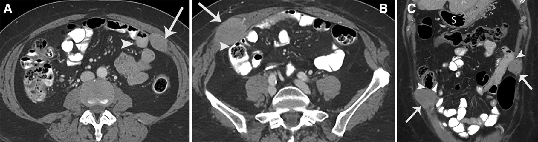
Figure 2A shows an enhacing mass in the left internal oblique muscle, and infiltrates into the external oblique muscle (arrow). The mass abuts the proximal ileal small bowel loop (arrow head). Figure 2B shows a mass in the right abdominal wall infiltrating the internal and external oblique muscle and the tranverses abdominus muscle (arrow). The mass abuts the adjacent colonic loop (arrow head). Figure 2C shows the two enhancing masses involving the abdominal wall (arrows) and abutting the adjacent bowel loops (arrow heads). Note there is no serosal involvement of the bowel loops. The stomach (S) is located superior to the transverse colonic loop.

(A) Axial post-contrast CT of the abdomen showing mass in the left internal oblique muscle. (B) Mass in right abdominal wall. (C) Coronal post-contrast image showing the two enhancing masses involving the abdominal wall.
These masses were suggestive of laparoscopic port implants and recurrent disease was confirmed by a ultrasound-guided biopsy in October 2019.
Dr Malpica
The metastatic tumor consists of a gland-forming neoplasm with squamous morules. Metastatic endometrial endometrioid carcinoma was shown in the abdominal wall. There was evidence of tumor fragments adjacent to fibroconnective tissue and the inset shows a higher magnification of the tumor (Figure 3). Inmunochemical stains showed that the neoplastic cells were positive for estrogen receptor and PAX8.

Metastatic endometrial endometrioid carcinoma in the abdominal wall, tumor fragments adjacent to fibroconnective tissue. Inset shows a higher magnification of the tumor.
DR MEYER: Given the multiple sites of recurrence at the port sites, what would be your recommendation for the patient at this time?
Factors that must be considered in this patient include the multifocal nature versus single port-site metastasis and the suspicion of involvement through the peritoneum on CT imaging. Options include primary surgical resection followed by chemotherapy or radiation, systemic treatment with standard chemotherapy or hormonal therapy, or consideration of clinical trials. Consultation with a plastic surgeon is recommended for planned abdominal wall reconstruction after surgical resection. Given the size and multifocal nature of the port-site metastases, and potential morbidity associated with resection, primary surgical resection may not be favored in this case. Previous data have shown that radiotherapy for isolated port-site metastases in endometrial cancer is associated with high rates of local control. 3 However, in this patient, radiation therapy may not be ideal given the size of the port-site metastases and the multifocal manifestation of the recurrence. In addition, concern that there could be bowel adherence to the intra-peritoneal component of the metastases precluded this option.
Hormonal therapy may also be considered. Data from a phase II study evaluating the mammalian target of rapamycin inhibitor, everolimus, in combination with letrozole, showed promising results in patients with recurrent endometrial cancer. The clinical benefit rate was 40% and the confirmed objective response rate was 32%. In that study, none of the patients discontinued treatment as a result of toxicity. 4 More recently, another phase II study evaluated the same combination but with the addition of metformin in patients with recurrent endometrioid endometrial cancer. The authors found a clinical benefit rate of 50% and a 28% objective response rate. 5
One should also consider molecular testing, and determine whether there is evidence of microsatellite instability or mismatch repair deficiency in such a patient. Recent data, from an open-label, single-arm, phase II study evaluated the combination of lenvatinib, a multikinase inhibitor of vascular endothelial growth factor, and other tyrosine kinases, and pembrolizumab, an antibody targeting PDL-1 in patients with metastatic endometrial cancer. 6 The investigators found an objective response rate of 39.6%. Only 9% of patients discontinued treatment due to adverse events. Therefore in this patient, consideration of systemic therapy would be most sensible. Additionally, molecular tumor testing, biomarker testing (PDL-1), and testing for microsatellite instability status could guide additional systemic therapy options.
Ultimately the patient elected to proceed with a clinical trial (MD Anderson Protocol 2015–0723) with carboplatin AUC 5, paclitaxel 175 mg/m2 IV, and enzalutamide 160 mg PO daily. The patient began her treatment in December 2019.
Dr Meyer
Closing summary
Port-site metastases are rare, with an estimated incidence of 0.18% to 0.33% in early-stage endometrial cancer. 7–9 Although the pathophysiology of port-site metastases is not fully understood, a variety of hypotheses, including the efflux of gas around the trochars, surgical technique, biologic properties of individual tumors, and the decreased immune response from smaller incisions, have been proposed. 10–14 Some easily implementable actions that may decrease the risk of port-site metastases include minimizing tissue trauma and the number of instrument transfers, rinsing trochars in 5% povidine-iodine before insertion, rinsing tips of instruments and irrigating port sites with 5% povidine-iodine, fixating trochars, placing all specimens in a bag before removing through trochars, removing intra-abdominal and pelvic fluid before removal of trochars, deflating the abdomen with trochars in situ, closing the peritoneal trochar sites of 10–12 mm trochars, and resecting tumors with adequate margins. 14 Other interventions studied in animal models include lavage of port sites with chemotherapy or heparin. 15
In conclusion, the data show a poor prognosis both for patients with non-isolated and isolated port-site metastases. Prognosis is probably poor because, regardless of the presentation, port-site recurrence may be a surrogate for a more aggressive tumor biology with disseminated and micrometastatic disease at the time of manifestation. We know that solitary port-site recurrences are rare in early-stage endometrial cancer. Given their rarity, data on the most effective treatment strategy are limited, although success has been reported with each of the three standard methods as well as combinations of surgical resection, radiation, and/or chemotherapy. Careful consideration of individual tumors and clinical characteristics should continue to guide therapeutic decisions.
References
Footnotes
Twitter @BSegarraVidal
Contributors BS identified the patient, and LAM manage treatment of the patient. AM and PB contributed with diagnostic investigations.
Funding The authors have not declared a specific grant for this research from any funding agency in the public, commercial or not-for-profit sectors.
Competing interests None declared.
Patient consent for publication Not required.
Provenance and peer review Commissioned; internally peer reviewed.