Article Text

Statistics from Altmetric.com
Case Presentation
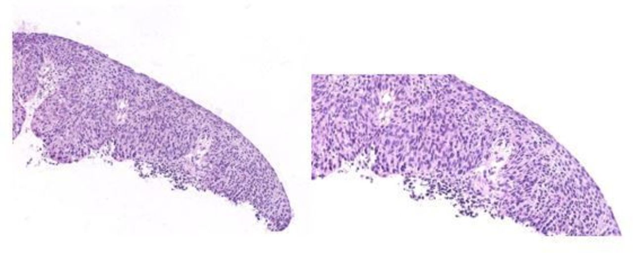
An asymptomatic 60-year-old woman, G2P0 (body mass index 23.14 kg/m2) with a past medical history significant for placement of a bypass valve for a cystic lesion in the third cerebral ventricle and also a history of secondary epilepsy. She had undergone a laparoscopic extrafascial hysterectomy and bilateral salpingectomy 14 months prior to consultation (November 2017). The indication for the hysterectomy was for cervical dysplasia. She was not considered a candidate for a third conization, given the scarce remaining cervical tissue and her age. The patient had two previous consecutive conizations showing cervical intra-epithelial neoplasia III (CIN III) with exocervical positive margins for dysplasia (Figure 1). The patient was then offered a simple hysterectomy. The final pathology on the hysterectomy specimen was significant for an isolated focus of CIN III, with a foci of signs of human papillomavirus (HPV) infection (visualization of koilocytosis: nuclear enlargement, hyperchromasia, and a perinuclear halo), with cervical margin free of any dysplastic lesions (Figure 2).

Conization. Detail of cervical mucosa with disordered cell proliferation showing the lack of maturation and loss of normal histological structure (dysplasia). The cells vary in size and have hyperpigmented atypical nuclei. This lesion reaches the outer third of the total thickness of the epithelium (cervical intra-epithelial neoplasia (CIN) III).

Detail of cervical mucosa, in hysterectomy specimen, where mature squamous epithelium of normal appearance (right side) and proliferated and atypical epithelium can be seen with increased core/cytoplasm ratio, nuclear hyperpigmentation, and lack of maturational order that reaches the superficial third of the total thickness of the epithelium (cervical intra-epithelial neoplasia (CIN) III).
Dr Baiocchi
How should one proceed in the setting of multiple conizations with positive margins for cervical dysplasia in a post-menopausal woman?
In the setting of multiple conizations for cervical dysplasia in a post-menopausal woman, a simple hysterectomy is the preferred procedure. As a cervical ablation is not recommended due to inadequate colposcopy and absence of the pathologic specimen, hysterectomy as a definite treatment is acceptable for patients when it is not feasible to repeat the diagnostic excisional procedure (conization). 1
One of the main concerns is the risk of an underlying invasive cervical cancer diagnosed after the hysterectomy. The incidence of occult invasive lesion in this setting is not clear. Mahnert et al 2 found a 2.5 % (5/196) risk of invasive cancer after hysterectomy for cervical dysplasia in a large cohort of 6360 patients who underwent hysterectomy for presumed benign conditions. However, up to 5% – 15 % of cervical cancer cases are diagnosed after an inadequate simple hysterectomy. 3 4 The most important causes of ‘ cut-through ’ hysterectomies are not only the absence of an invasive lesion in the conization specimen, but also a false-negative Pap smear and inadequate evaluation of colposcopy.
Nevertheless, the presence of positive margins after conization is a relatively common finding and highly related to an increased risk of recurrence of dysplasia. In a recent meta-analysis by Arbyn et al,5 positive margins were described in 23.1 % of cases. The overall risk of recurrent CIN II + was 6.6 % and relative risk increased by 4.8-fold in positive margins compared with negative margins. Moreover, the risk of recurrence was only 3.7 % for free margins and even lower (0.8%) with a post-treatment negative HPV test.
After the hysterectomy, what would be your routine surveillance recommendations?
Although there is convincing evidence that screening women who have had a hysterectomy with removal of the cervix for indications other than a high-grade pre-cancerous lesion or cervical cancer provides no benefit, 6 there is no established guideline suggesting the surveillance for those patients that had hysterectomy and previous history of cervical dysplasia.
For women treated for CIN II+, the current guideline published in 2012 1 recommends co-testing at 12 months and 24 months . If both co-tests are negative, is recommended to retest in 3 years . If all tests are negative, routine screening is recommended for at least 20 years , even if this extends screening beyond 65 years of age. This guideline may be extrapolated and applied for patients that had hysterectomy for cervical dysplasia. Nevertheless, few studies addressed the surveillance of women with CIN at hysterectomy. Gemmel et al 7 examined 341 women who underwent hysterectomy for CIN III and 219 completed a 10- year follow-up. Only eight (4%) presented with abnormal cytologies – six cases following normal cytologies, only two (0.91%) had persistent vaginal intra-epithelial neoplasia ( VAIN ) and no patient developed vaginal cancer. Wiener et al 8 studied 193 patients – 143 patients completed a 10 - year follow-up and 43 completed a 2 0- year follow-up . Five patients presented with abnormal vaginal cytologies (VAIN 2 and invasive vaginal cancer), three within the first 20 months after hysterectomy. One invasive vaginal cancer was diagnosed 16 years after hysterectomy. A cumulative incidence of VAIN after hysterectomy was estimated to be 0 % at 10 years, 0.8 % at 15 years , and 2 % at 20 years . Kalogirou et al 9 reported 993 patients who underwent hysterectomy for CIN III – 793 cases with 10- year follow-up. They identified 41 VAIN cases (5.1%) – four patients had VAIN 1 and 37 patients had VAIN 2+. Most VAIN cases developed in the first 2 years of follow-up. Finally, Schockaert et al 10 found in 94 women (including stage Ia1 cervical cancer), seven (7.4%) who developed VAIN 2+, of which two were invasive vaginal cancers. Median interval between hysterectomy and diagnosis of VAIN 2 + was 35 months . Overall, in these four series, a total of 1249 patients had at least 10 years of follow-up and only 48 (3.8%) patients presented high-grade VAIN and three (0.2%) patients with invasive carcinomas.
Other large studies have documented the overall low risk for abnormal cytology after hysterectomy. A retrospective cohort study included 5862 women after hysterectomy for benign disease and found abnormal vaginal cuff cytology among only 79 (1.3%) women, with a mean time of 19 years . 11 Moreover, a cross-sectional study included 5330 screening cytology tests in women after a hysterectomy and reported only one case of dysplasia and no cases of cancer. 12 Therefore, even for women that have an increased risk of vaginal cancer due to hysterectomy after CIN, there is no established method to effectively impact the development of this rare condition.
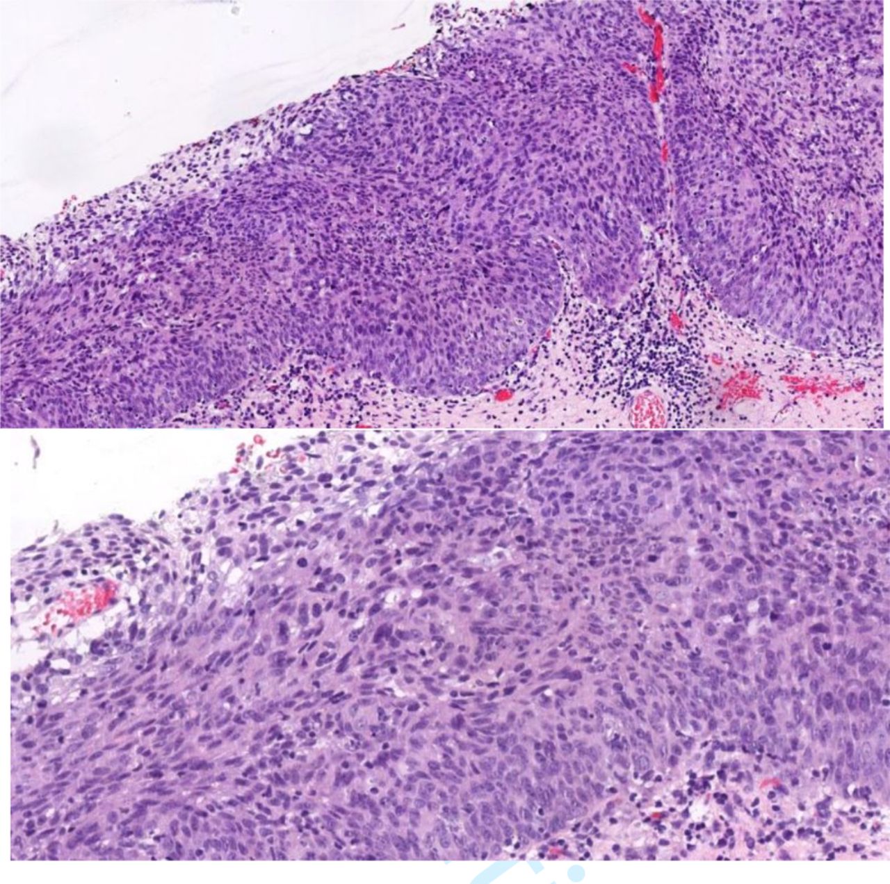
The following year (November 2018), during a routine surveillance visit, a Pap smear of the vagina was performed with a diagnosis of high-grade squamous intra-epithelial lesion (HSIL) (HPV16+). In our institution, after conization, patients are recommended to have a liquid-based Pap smear and HPV genotyping twice per year for the first the year (6 months apart) and then yearly, or yearly after hysterectomy. The patient underwent colposcopic evaluation of the vagina that showed a raised lesion at 3 to 6 o’clock at the apex of the vagina in the region of the colpotomy. The lesion measured approximately 2 cm. Under acetic acid a mosaic area with increased vascularization was visualized. The Schiller test showed an area of concern after application of Lugol’s solution. A biopsy was performed and the findings revealed VAIN 2–3 (Figure 3).
Vaginal biopsy. Detail of vaginal mucosa with disordered cell proliferation (dysplasia), increased nucleus/cytoplasm ratio, and marked nuclear atypia with uneven sizes and dark coloration. These alterations reach the superficial third of the epithelium (vaginal intra-epithelial neoplasia (VAIN) 3), while the more superficial cell layers maintain a normal disposition.
Dr Baiocchi
At this point, would you provide any other options than surgical excision of the lesion?
The incidence of VAIN is 0.2 – 0.3 per 100 000 women. 13 It comprises 0.5 % of all neoplastic lower genital tract lesions and 1 % of all intra-epithelial neoplasias. 13 Although there have been several published studies, lack of evidence on the management of VAIN exists, as most series are retrospective and include a small number of cases. Treatments include interventional or conservative modalities such as surgical resection, radiotherapy, ablation, and medical management. The treatment choice is usually based on the characteristics of the lesion (number of lesions, grade, location, and suspicious of invasion) and of the patient (age, previous radiation therapy, previous treatments, and sexual activity).
Traditionally, partial or total vaginectomy and radiotherapy were considered the only choices for high-grade VAIN treatment. 13–16 However, both treatments cause severe adverse effects that may negatively impact quality of life. Considering that VAIN are now mostly diagnosed in younger women, conservative approaches are preferable. In young women with multiple lesions, the treatment of choice may be CO2 laser ablation. 17–19 It has the advantage that can be repeated several times and with a success rate of nearly 70% – 80 % . 19 20 However, partial upper vaginectomy should still be the treatment of choice for apical VAIN 3 or VAIN in the region of the vaginal cuff scar in women hysterectomized due to cervical neoplasia. 13 Upper vaginectomy provides a histopathologic diagnosis and it has a good success rates. 21 22
The main concern of conservative management is missing a vaginal cancer. Therefore, excisional procedures should be preferred in the presence of any suspicious of invasive disease. Recently, Cong et al 23 analy zed 86 cases that underwent upper vaginectomy after a diagnosis of vulval intra-epithelial neoplasia 2 + (VIN 2 + ) and found a high rate (18.6%) of invasive disease. Fifty-six cases had previous hysterectomy due to HSIL and five (8.9%) with invasive disease. Moreover, 30.2 % of cases with suspicious features of invasion in colposcopy (atypical vessels, fragile vessels, irregular surface, exophytic lesion, necrosis, ulceration) had invasive disease.
Few studies compared treatment modalities of high-grade VAIN. Bogani et al 19 compared 169 (82.8%) patients who underwent laser ablation for VAIN to 35 (17.2%) who underent excisional procedures. A total of 41 (20%) patients developed high-grade VAIN at a median follow-up of 65 months . Only HPV persistence (HR 2.37) was associated with the risk of recurrence at multivariate analysis. Seven (3.4%) invasive cancers were observed. The type of procedure did not influence persistence of HPV infection and recurrence rates were similar between groups. Recently, the same group 18 analyz ed 117 cases of recurrent (median time of 20 months ) high-grade VAIN that had laser ablation or medical treatments. Patients with recurrent high-grade VAIN undergoing medical treatments were at higher risk of developing a second recurrence compared with women having laser treatment ( p = 0.013 ).
Another recent large series of 576 cases by Kim et al 24 found that laser or excision provides higher regression rate than topical agent or observation, and high-risk HPV positivity is a risk factor for recurrence. Patients with VAIN are at high risk of recurrence, but the risk of progression to vaginal cancer was relatively low. In VAIN 2 + patients , management included mostly topical management, laser ablation, and excision with recurrence rates of 62.5%, 26.4 % and 32.7 %, respectively. Four (3.2%) patients developed invasive disease with a median time follow-up of only 21.4 months .
In summary, laser ablation should be the preferred conservative treatment, especially in multifocal and young women. Any suspicious lesion should be excised for pathologic assessment and upper vaginectomy should be considered after hysterectomy, especially when an adequate colposcopy is not feasible.
What is the role of radiotherapy in this setting? Please speak specifically about external radiation and brachytherapy?
Few studies of intracavitary brachytherapy for VAIN treatment have been reported including both low-dose rate ( LDR ) and high-dose rate ( HDR ) modalities. However, brachytherapy has proven its effectiveness in the treatment of VAIN. 16 25–31 Additionally, external beam radiotherapy has also been described. However, the low rate ( < 1% ) of pelvic lymph node involvement is probably related to an undiagnosed invasive disease. Therefore, external beam pelvic radiotherapy is unwarranted as pelvic or distant recurrences after brachytherapy have not been observed in the absence of the invasive disease. 16
Although there are no studies comparing LDR to HDR regarding outcomes and toxicity, HDR has been the first option in recent years. 28 32 Notably, the variety of total prescribed doses shows a lack of the recommended method as the administered doses ranges between 40 and 70 Gy in both HDR and LDR with a prescribed depth of 0 to 1 cm . 16 25–32 Dose clearly affects toxicity, as ≥ 70 Gy had a 71.4 % of severe vagina complication rate, un like lower doses. 25 Moreover, patients treated with 60 Gy with a distance of 0.5 cm from the applicator had a low toxicity profile. 16
The relapse rate after brachytherapy seems to be lower or even similar to other treatment modalities, ranging from 0 % to 14 % . 13 16 25–32 The main question is whether brachytherapy in high-grade VAIN is sufficient to treat occult invasion, and the major criticism compared with surgery is the absence of a pathologic specimen for detailed diagnosis. Another concern is the potential adverse impact of brachytherapy in future surgical procedures and the risk of a radiation-induced secondary neoplasm. Moreover, radiotherapy itself may increase the development of VAIN, found in about 20 % of women that received radiotherapy for gynecologic malignancies. Furthermore, VAIN in women with previous radiotherapy seems to be more refractory to treatment and more likely to invasive cancer progression. 33
Nevertheless, few studies evaluated the sexual function after brachytherapy for high-grade VAIN. Woodman et al 26 reported satisfactory intercourse in 9/10 cases, despite the entire vagina radiation. In a series of 22 patients , Graham et al 30 reported late vaginal mucosal changes such as atrophy, vaginal dryness, telangiectasia, stenosis, ulcers, and also sexual dysfunction due to vaginal dryness and dyspareunia. 30 Recently, 3D planned imaging permits a volumetric distribution of the dose to the target volume with preservation of surrounding organs with better outcome results and lower toxicity. Moreover, the irradiated vagina should include only the VAIN lesions, forgoing the treatment of the entire vaginal length.
In summary, brachytherapy has been shown to be an effective method of VAIN treatment and recent brachytherapy techniques should help to mitigate toxicity. The risk of severe toxicity is low but patients must be counseled appropriately regarding the likelihood of mild to moderate toxicity, including sexual dysfunction. Radiotherapy should not be recommended as a first-line treatment of high-grade VAIN but may need to be considered for cases that are resistant to other conservative and surgical treatment. Radiotherapy has its own implications in terms of early and late adverse effects, impact on sexual function, and risk of developing vaginal malignancy.
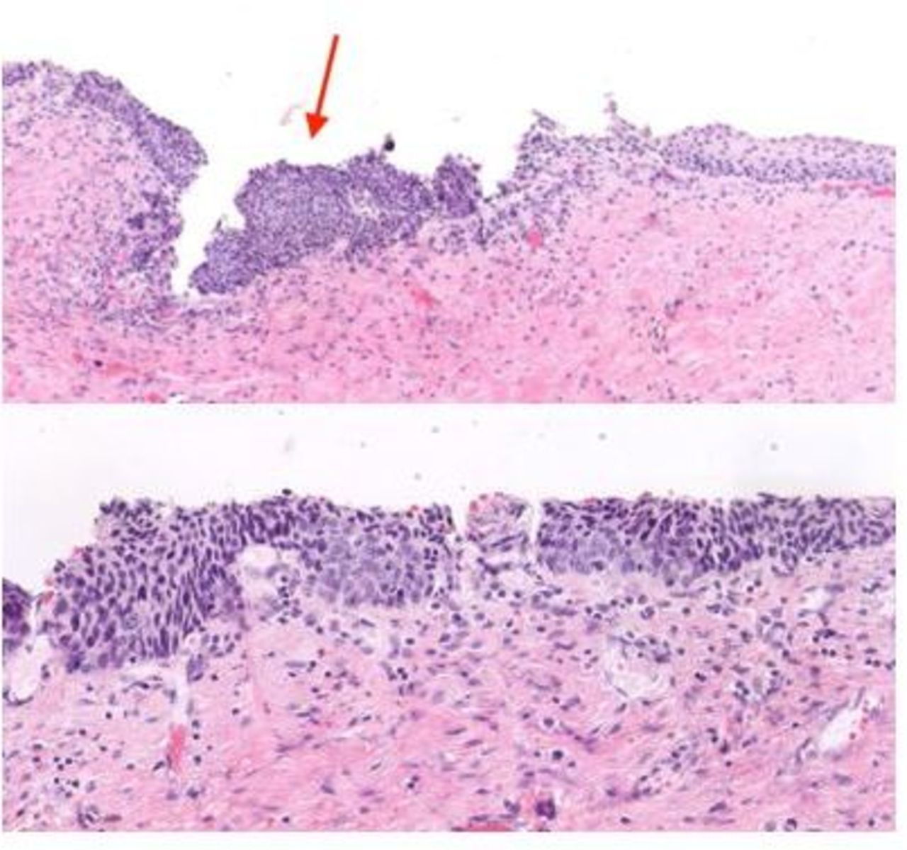
The patient was then scheduled on January 2019 for surgical excision of the vaginal lesion, performed via a vaginal approach. The final diagnosis was a moderately differentiated infiltrating carcinoma, with stromal invasion of 4 mm in contact with the deep margin and horizontal extension of 8 mm, without lymphovascular invasion (Figure 4A–D).

Surgical vaginal resection histology slides: A: Neoplastic proliferation of squamous epithelial cells is observed, infiltrating the stroma. Nests and irregular islands of atypical cells are observed, not ordered according to the normal histologic arrangement of healthy tissue, embedded in a desmoplasic stroma with associated inflammation Infiltrating lesion that contacts the margin of resection marked with Chinese ink. B: Polygonal and fusiform cells with variable eosinophilic cytoplasm, oval, or rounded nuclei with a prominent nucleolus and a varied degree of pleomorphism are observed. Isolated dyskeratotic cells and weak intercellular bridges are observed. Mitosis figures are frequent. C: Infiltrating lesion of squamous epithelial cells, positive expression with keratin CK5/6. D: The association with human papillomavirus is related to the positivity of the p16 immunohistochemical marker (positive staining in the infiltrating lesion: intense and diffuse color) which, however, is negative in normal tissue.
Dr Juaristi
Neoplastic proliferation of squamous epithelial cells is observed, infiltrating the stroma and contacting the margin of resection. The cells are polygonal and fusiform with variable eosinophilic cytoplasm, oval or rounded nuclei with a prominent nucleolus, and a varied degree of pleomorphism. Isolated dyskeratotic cells and weak intercellular bridges are observed. Mitotic figures are frequent. The infiltrating lesion of squamous epithelial cells was positive expression with keratin CK5/6. The association with HPV virus can be seen in the infiltrating lesion with immunohistochemical marker p16, which is negative in normal tissue.
Dr Baiocchi
Please provide details as to what should be the recommended work-up for a patient with this diagnosis before proceeding with further treatment recommendations?
Vaginal cancers are characterized for local invasion and disseminate by several routes. Direct extension occurs to surrounding structures such as paravaginal tissues, paracervix, bladder, urethra, vulva, and rectum. Lymphatic drainage of the vagina is complex, with an extensive communicating network of the submucosa and muscularis that follows the course of uterine vessels in the upper third of the vagina and the vaginal vessels in the lower two-thirds. Lymphatic dissemination of the upper third usually occurs first to the pelvic lymph nodes and rarely to para-aortic lymph nodes. The lower third of the vagina disseminates mainly to the groin nodes, but also to femoral lymph nodes. Tumors located in the middle third may follow both lymphatic routes.
Careful vaginal examination and total inspection of vaginal walls must be carried out. Any suspicious lesion should be biopsied and it may present as a tumor, ulcer, or even a plaque. In the absence of a macroscopic lesion and abnormal Pap smear, a colposcopy must be performed. Sometimes multiple biopsies from various sites need to be accomplished to ensure invasion. Local primary disease within the vagina, size, and macroscopic characteristic (ulcerative or exophytic) should be assessed careful by gynecologic physical exam, and suspicious inguinal lymph nodes should be described.
Despite recent update on International Federation of Gynecology and Obstetrics (FIGO) cervical cancer staging, 34 for vaginal cancer the important prognostic factors such as lymph node involvement and advanced imaging modalities may not be used to change the tumor staging. Staging for vaginal cancer is still based on clinical evaluation according to the 2009 FIGO system. This system allows only basic work-up such as chest radiography, gynecologic examination (bimanual and rectovaginal examination), cystoscopy and/or proctoscopy, and intravenous pyelogram. However, FIGO encourages the use of other and some advanced imaging modalities, such as computed tomography (CT), magnetic resonance imaging (MRI) , and positron emission tomography ( PET ) to guide management.
MRI is particularly useful for tumor size and locoregional evaluation. It is more sensitive than physical examination in assessing paravaginal or parametrial involvement in women with cervical cancer, and that might be similar to vaginal cancer. 35–37 Vaginal tumors are best visualized on T2-weighted images and are characterized as low-to-intermediate signal intensity on T1-weighted imaging and intermediate-to-high signal intensity on T2-weighted imaging. 35 38 39 The vaginal walls may be distended by vaginal gel that allows better assessment of the tumor details and surrounding tissues. 35 38 39 As the tumor is limited to the vagina in stage I, the paravaginal fat remains in high signal intensity on T1-weighted images. Conversely, in stage II tumors, the normal low signal intensity vaginal wall cannot be identified, and the paravaginal fat is of abnormal low signal intensity on T1. Moreover, MRI has an accuracy rate of 92 % for all metastatic tumors involving the vagina. 35 38 39
Data on the role of fluorodeoxyglucose (FDG)-PET imaging in the management of vaginal cancer are relatively scarce and usually described along with other gynecologic tumors. Similarly to cervical cancer, it appears to have better sensitivity than other modalities in detecting metastatic inguinal or pelvic lymph nodes. 40 41 In a series by Robertson et al, 41 29 patients with vaginal cancer were included, and PET-CT changed the physician management in 13 (45%) cases. Moreover, CT can also be used to evaluate nodal and distant metastatic spread, and rectosigmoidoscopy or cystoscopy should be performed when there is a clinical or radiologic suspicious involvement of the urethra, bladder, anus, or rectum. 35
In summary, physical examination and pelvic MRI are of great value in vaginal cancer work-up. Based on the suspicious or higher risk of distant metastasis, especially in stage ≥II, a PET-CT should be used if available. CT scan may also assess distant metastasis in the absence of PET-CT. In the absence of CT scan, abdominal ultrasound and chest radiography should be used. Furthermore, I might agree with the work-up performed by the authors, after the diagnosis of an early-stage disease.
What should be the recommendation for this patient with positive margins in the vaginal resection specimen?
As the lesion involves the upper third of the vagina and no adverse risk factors that indicate radiation therapy were present, surgery including colpectomy and lymph node dissection may be the treatment choice. This recommendation is similar to cervical cancer management after an inadvertent simple hysterectomy. In both scenarios, patients who may benefit from surgery are those without adverse risk factors that already indicate radiation therapy. However, in vaginal cancer no data exist regarding the adequate margins or how radical should be the paravaginal resection. For stage I vaginal carcinoma, in most cases a type B resection or type C1 in case of larger tumors (>2 cm) may be adequate for clear margins. Yet, radiation therapy would be an option for this particular case if surgery were not feasible for other reasons.
Stage I vaginal carcinoma can be adequately treated with radiotherapy, with or without surgery, depending on the tumor site. Lymph node metastasis is relatively uncommon, and estimated at from 6% to 16 % . 42–44 Due to its rarity, the surgical procedure has not been well standardized, and several different approaches have been reported. In selected patients with small tumors, wide local excision with disease-free margins has been adopted. 44–47 Moreover, partial vaginectomy, 44 total simple vaginectomy, 45–47 and also radical vaginectomy with paravaginal tissue resection have also been described. 44–46 Indication of surgery for stage I vaginal cancer should have risks and benefits balanced, and be restricted to young patients with tumors located in upper third and without previous indication of radiotherapy for adverse factors ( eg, presence of positive nodes). In the literature, eight series that included 174 patients showed a 5- year survival of 56%– 90 % in stage I disease treated with surgery alone. 48 For stages ≥ II , treatment should be based on radiation therapy and pelvic exenteration should be restricted to locoregional persistence or recurrence after radiotherapy.
Radiotherapy alone may also be used for stage I treatment as 22 studies that analyzed a total of 553 patients have reported a 5- year survival of 33 %− 100 % . The most commonly adopted strategy is a combination of interstitial or intracavitary therapy with external beam radiotherapy in patients with high-risk prognostic factors. 48 External beam radiotherapy is generally advisable for larger, more infiltrating or poorly differentiated tumors that may have a higher risk of lymph node metastasis. 49 50 Notably, a National Cancer Database ( NCDB ) analysis compared surgery and radiotherapy in women affected by stage I vaginal cancer and reported a better survival for surgery compared with radiotherapy, with a 5- year survival of 90 % versus 63 % ( p < 0.05 ). 51
The adoption of chemotherapy added to radiotherapy in vaginal cancer followed the experience of cervical cancer treatment. An analysis of the NCDB showed a clear benefit in favor of chemoradiation compared with radiotherapy alone for all stages. In stage I tumors, the median survival was 85.3 versus 109 months ( p = 0.02 ). 52 Tumor size also impacts survival in stage I patients . For the 293 stage I patients from the Surveillance, Epidemiology, and End Results (SEER) database, the 5- year survival for tumor size ≤ 2 cm and > 2 cm were 79.2 % and 66.1 % , respectively ( p = 0.01 ). 53
In summary, the primary treatment options in cases of vaginal carcinoma limited to the vaginal mucosa are surgery or radiotherapy. For tumors located in the upper third of the vagina, surgery may be preferred. Radiotherapy can also be used with a curative intent in stage I, with a minimum brachytherapy dose of 75 Gy. External beam radiotherapy and chemotherapy should be added to brachytherapy as they would improve local control and survival rates, especially in high-risk disease.
Given this finding, a pelvic MRI and a chest X-ray was requested and this revealed no evidence of residual disease or adenopathy. At a multidisciplinary tumor board, it was decided to perform a laparoscopic bilateral pelvic lymphadenectomy. A total of 13 lymph nodes were removed, six on the right and seven on the left side, all were analyzed and were negative on intra-operative frozen section and on final diagnosis. The frozen section evaluation was performed since if these lymph nodes were found to be positive for disease, the procedure would have been aborted and a recommendation for concomitant radiation and chemotherapy implemented. All findings revealed no evidence of disease. The patient also underwent a laparoscopic radical parametrectomy (type B excision), including an upper vaginectomy. The final pathology revealed no evidence of cancer or dysplasia in the radical parametrectomy specimen.
Dr Baiocchi
What is the role of lymphadenectomy for a patient with these findings and is there a role for sentinel lymph node mapping?
Lymph node metastasis in stage I vaginal cancer ranges from 6% to 16 % . 42–44 Due to its rarity, to date only four published articles have addressed the value of sentinel lymph node ( SLN ) mapping in vaginal carcinoma, comprising a total of 16 cases in stages I and II. 54–57 Notably, the overall detection rate was 81 % (13/16 cases). The largest series by Frumovitz et al 54 included 14 cases ( seven squamous cell carcinomas) and found that the lymphatic drainage did not follow the lymphatic channels that would have been predicted anatomically, whereas 1/3 of cases treated with radiotherapy had their field altered as result of the lymphoscintigraphy findings. Despite its rarity, the drainage for unexpected sites is an important argument to consider SLN mapping and further studies are needed to clarify its role in vaginal cancer.
Please provide your thoughts with regard to the accuracy of frozen section evaluation of the pelvic lymph nodes in this setting?
There are no data reporting the potential role of lymph node frozen sections in vaginal cancer. However, an analogy for vaginal cancer should be done considering the current data of frozen sections of SLN in cervical cancer. In early-stage cervical cancer management, one key objective is the avoidance of the potential morbidity related to combined radical surgery followed by pelvic radiotherapy. Therefore, an intra-operative SLN evaluation may be done, and in the case of SLN involvement the radical surgical procedure should be abandoned and the patient referred for chemoradiation.
However, published series have described a low sensitivity of intra-operative SLN frozen section, especially for low-volume metastasis. 58–60 Bats et al 58 reported the data from the SENTICOL trial (Ganglion Sentinelle dans le Cancer du Col), and failed to detect node involvement in 15/20 cases (three cases of macrometastasis). Roy et al 59 evaluated 211 patients , 10/13 cases with positive SLNs were false-negative by frozen section, including one macrometastasis. Slama et al 60 corroborated this finding in 225 patients , where intra-operative SLN evaluation correctly detected only 39/73 cases (53%) with positive SLNs and missed eight cases of macrometastasis. Conversely, Martinez et al 61 found a better sensitivity (89%) of frozen section in a smaller study that included 94 patients .
In summary, frozen section of SLNs in cervical cancer is limited by the high false-negative rate. However, intra-operative SLN analysis results in the detection of 50% of the cases with node involvement and detects most macrometastasis. Finally, this data may be extrapolated and somehow applied in vaginal cancer management.
If this patient would have had evidence of positive pelvic lymph nodes, would there have been a role for para-aortic lymphadenectomy?
The exact para-aortic lymph node involvement rates and its prognostic value in vaginal carcinoma have not been described. However, the data from cervical cancer management may be applied. In locally advanced cervical cancer, 22 % of para-aortic positive nodes are expected in cases of pelvic positive nodes 62 and surgical para-aortic staging modifies treatment in 18% – 33 % of cases a s it indicates extended field radiation. 62 63 Moreover, recent data from a phase III trial (Uterus-11) showed that laparoscopic para-aortic staging might also impact survival. 64
In summary, no data support para-aortic staging in vaginal cancer for tailoring extended field radiation. Moreover, the role of para-aortic lymph nodes as a site of recurrence is not clear for vaginal cancer. Although data from cervical cancer may be extrapolated, it should not be advised as a standard treatment for vaginal carcinoma.
Is there any role for complete resection of the vagina?
Despite the need for close surveillance and the concern about recurrence during follow-up, one of the major benefits of surgery instead of radiation therapy is to decrease late morbidity, maintenance of vaginal length, and also sexual function. Moreover, total vaginectomy theoretically increases the surgical morbidity, where the risks may not overcome the potential benefit of complete resection.
In May 2019, the patient was evaluated for routine surveillance. In our department, we follow the surveillance recommendations of the European Society of Gynecological Oncology (ESGO) guidelines for cervical cancer. We perform a physical (rectovaginal and abdominal) examination, evaluation of inguinal and supraclavicular areas, and a liquid-based cytology every 3 months during the first 2 years, every 6 months for the next 3 years, and then annually thereafter or until year 10 or discharge at the discretion of the treating physician. We also perform a chest X-ray and tumor marker (squamous cell carcinoma) annually. Although squamous cell carcinoma antigen has not been proven to be useful in vaginal cancer, it may be increased in patients with epidermoid carcinoma of the cervix, benign tumors of epithelial origin, and benign skin disorders, and may be helpful in determining relapse when monitoring patients with complete remission in cervical cancer. The patient had recovered well from surgery and had no complications.
Dr Baiocchi
Closing Summary
Please give us some information about progression rates from VAIN 2 – 3 to vaginal cancer
HPV plays a central role in the etiology of VAIN, similar to that of CIN and VIN. 65 66 HPV were found in low-grade VAIN in 98% – 100 % of cases, 90% – 92.5 % in VAIN 2 – 3 and in 65% – 70 % of invasive carcinomas. HPV 16 and 18 were found in 64 % of VAIN 2 – 3 cases and in 72 % of invasive lesions. 13 67 68 The precise natural history of VAIN is still unclear and the potential VAIN progression into cancer is not precisely understood. Observational studies have shown that 90 % of low-grade VAIN probably regress spontaneously. 20 69 For the minority of VAIN 1 c ases that progress to VAIN 2 – 3 , an interval of nearly 15 years has been described. 70 Further VAIN 3 progression rates to invasive carcinoma after adequate treatment ranges from 2 % to 5 % . 13 71 72 VAIN is located at the upper third of the vagina in 80 % of cases, multifocal in up to 60 % of cases, and mostly found in women after hysterectomy for CIN2+ (80%). 3 20–22 70
Unfortunately, even after appropriate treatment, VAIN recurrence occurs in one-third of cases . 13 71 72 Treatment of VAIN comprises interventional or conservative modalities such as surgical resection, radiotherapy, ablation, and medical management. The treatment is chosen based on the characteristics of the lesion (number of lesions, their grade, location, suspicious of invasion) and of the patient (age, previous radiation therapy, previous treatments, and sexual activity).
In young women and multifocal disease, conservative management with laser ablation should be chosen. In cases of upper vault lesions with difficult access to colposcopy or any suspicious of invasion, an upper vaginectomy or local excision is preferred. Brachytherapy is an efficacious method, but should not be recommended as a first-line treatment of high-grade VAIN due to the late toxicity profile. It should be considered for cases that are resistant to other conservative and surgical treatments.
In cases of vaginal carcinoma, the treatment is defined by primary tumor location and stage. MRI is the best imaging for locoregional evaluation and PET-CT has the best sensitivity for nodal and distant disease work-up, mainly for locally advanced tumors. For stage I tumors located in the upper third of the vagina, radical surgery with node staging or radiotherapy (external beam radiotherapy and brachytherapy) are the treatments of choice. For the middle and lower thirds of the vagina or stage ≥II, radiotherapy (external and brachytherapy – intracavitary or interstitial depending on the tumor burden) should be the preferred treatment. Radical surgery (such as pelvic exenteration) may be used in cases of recurrence or persistence after radiation therapy. Chemotherapy should be added to radiation even in stage I and SLN mapping may help to identify unexpected lymphatic drainage. Finally, I would like to congratulate the authors for this interesting and well-documented case.
References
Footnotes
Twitter @mgorostidi, @glaucobaiocchi
Contributors MG: idea, writer, review, and invitation. AL: review, supervision. AJ: pathologist for the case. GB: discussant, writer.
Funding The authors have not declared a specific grant for this research from any funding agency in the public, commercial or not-for-profit sectors.
Competing interests None declared.
Patient consent for publication Not required.
Provenance and peer review Commissioned; internally peer reviewed.