Article Text

Abstract
Background Invasive vulvar Paget’s disease with over-expression of the human epidermal growth factor receptor 2 (HER2) protein is potentially suitable for targeted therapy, especially in a metastatic setting where no effective treatments are available.
Methods Four consecutive patients with HER2 positive advanced vulvar Paget’s disease, treated with weekly trastuzumab (loading dose 4 mg/kg, then 2 mg/kg) and paclitaxel (80 mg/m2) followed by 3-weekly trastuzumab maintenance (6 mg/kg), are reported.
Results Median age and follow-up of patients were 62.5 years (45–74) and 16 months (6-54), respectively. Complete or partial responses were observed in all patients. Median time to response was 3 months (range 2–4), while median duration of response was 10 months (range 2–34). Case 1 presented with pulmonary and lymph nodes involvement. She experienced a radiological complete response after 24 treatment administrations, and a progression-free survival of 36 months. At disease progression, treatment re-challenge achieved partial response. She is currently receiving treatment with trastuzumab–emtansine. Case 2 was a 74-year-old woman who developed pulmonary metastasis after first-line cisplatin treatment. She had a partial response and a progression-free survival of 10 months. Case 3 had inguinal and para-aortic lymphadenopathy in complete response after 18 treatment administrations. She developed brain metastasis while receiving trastuzumab maintenance. Case 4 was treated for locally advanced disease and experienced a subjective benefit with relief in perineal pain and itching. No unexpected treatment-related side effects were reported.
Conclusions Advanced vulvar Paget’s disease is a rare disorder and no standard treatment is available. In the sub-group of HER2 positive disease, weekly paclitaxel–trastuzumab appears to be active and safe, and may be considered a therapeutic option in these patients.
- paget disease
- extramammary
Statistics from Altmetric.com
HIGHLIGHTS
Oncogene addicted advanced Paget’s disease could benefit from targeted anti-HER2 agents.
Single cases of vulvar Paget’s disease treated with a trastuzumab-based therapy have been previously reported.
This is the largest series of advanced vulvar Paget’s disease effectively treated with paclitaxel–trastuzumab.
INTRODUCTION
Extramammary Paget’s disease is a rare skin disorder that represents about 7–10% of cases of Paget’s disease.1 It is an epithelial neoplasia originating from the apocrine and eccrine glands of the epithelium, and is characterized by large cells (Paget cells) with prominent cytoplasm. In most cases, it localizes to the axillary or genital regions, and the vulva is involved in approximately 65% of cases.2 About 25% of invasive vulvar Paget’s disease over-expresses the human epidermal growth factor receptor 2 (HER2) protein on the cancer cell surface, unveiled by immunohistochemistry.3
Thus far, four case reports have described the successful use of trastuzumab-based therapies in patients with advanced HER2-positive vulvar Paget’s disease, for which previous extensive surgery was ineffective and no other therapeutic options were available.4–7 Here, we report four cases of advanced HER2-positive vulvar Paget’s disease effectively treated with weekly paclitaxel plus trastuzumab. Each patient was counseled about a lack of conventional treatment options and consented to trastuzumab–paclitaxel off-label therapy on the basis of previous case reports showing its efficacy and safety (online supplemental table). They were also informed about the large body of evidence in the use of this regimen for the treatment of HER2 positive breast cancer.
Supplemental material
METHODS
From 2012 to date, seven patients presented with an invasive recurrence of vulvar Paget’s disease, of whom four tested HER2 positive and are here reported. Summary statistics were used to summarize the observations. Median follow-up was measured since the first trastuzumab–paclitaxel administration, as for the other endpoints of efficacy.
RESULTS
Median age of patients was 62.5 years (range 45–74). Three patients presented with stage IV disease, and the last patient with locally advanced disease. Median progression-free survival was 13 months (range 6–36). Regarding toxicity and safety, paclitaxel-induced neuropathy was recorded in all four cases, along with alopecia and nail changes (dyschromia). Case 1 also had grade 3 neutropenia. Cardiologic assessment was performed at baseline and every 3 months during therapy, with no findings of cardiac dysfunction.
Case 1
A 45-year-old woman presented in October 2015 with a 16- year history of intra-epithelial Paget's disease of the vulva and anal/perineal region, with an underlying adenocarcinomatous component infiltrating the reticular dermis. She had previously received laser therapy, radiotherapy, imiquimod therapy, and photodynamic therapy in other hospitals for the intra-epithelial disease (Table 1). In February 2015, she experienced an infiltrative locoregional recurrence, with the need of a wide vulvo-perianal surgical resection, left colostomy and bilateral ilio-inguinal lymphadenectomy. Adjuvant radiotherapy was not proposed since lymph nodes were not involved. In October 2015, the patient had a new vulvar erosion attributed to local recurrence of invasive vulvar Paget’s disease. CT showed concomitant pulmonary involvement, including a 16 mm lesion in the right phrenicocostal sinus (Figure 1A), and a 14 mm lesion in the lower left lobe, as well as abdominal lymphadenopathies with one para-aortic lymph node of 25×22 mm and two iliac lymph nodes of 26×22 and 20 mm. Considering the patient’s performance status, age, previous treatments, and the HER2 over-expression unveiled on the pathological tissue resected in February 2015, we prescribed weekly paclitaxel 80 mg/m2 and trastuzumab 2 mg/kg (loading dose 4 mg/kg). The patient started first-line combination therapy on November 2015. The first CT in January 2016 showed a significant size reduction of the pulmonary nodules bilaterally, and the large lesion was no longer recognizable (Figure 1B). CT also showed a normalization in the diameter of the para-aortic and iliac lymph node. Even the vulvar lesion was no longer recognizable at clinical examination. She continued the combination treatment until July 2016, when she had a second scan confirming a complete response. During treatment, grade 3 neutropenia was registered, with several treatment delays and a dose reduction (80%) of paclitaxel from May 2016. Considering the hematological toxicity and the radiological complete response, maintenance monotherapy with 3-weekly trastuzumab (6 mg/kg) was started in August 2016; no other adverse events were reported. In August 2017 the patient was still in complete response and requested that the treatment be stopped. Therefore, a program of regular clinical and radiological follow-up was started.

(A,B) Case 1. Baseline Chest CT in October 2015 shows 16 mm pulmonary lesion in the right phrenicocostal sinus (A). Chest CT in January 2016 shows that the lesion is no longer measurable (B).
Summary of HER2 positive cases of vulvar Paget’s disease reported
CT re-assessment in November 2018 showed a 21 mm nodule in the perirectal region (confirmed cytologically) and new sub-pleural nodules in the lungs bilaterally. Considering the absence of residual toxicity and complete response to the previous treatment, a re-challenge with weekly paclitaxel–trastuzumab was started. CT revaluation in March 2019 showed a new radiological partial response, with the perirectal lesion no longer measurable and the sub-pleural lesions nearly disappeared. This result was confirmed in June 2019. The patient continued maintenance treatment with 3-weekly trastuzumab until January 2020, when the CT showed the re-appearance of the perirectal lesion and new pleural lesions and she reported worsening pain. Therefore, third-line therapy with 3-weekly trastuzumab emtansine 3.6 mg/kg was started in February 2020. At the first radiological evaluation on April 2020, CT showed a decrease in size of the perirectal lesion (from 38×29 mm to 25×17 mm). The patient is still receiving treatment with trastuzumab emtansine, with clinical benefit.
Case 2
A 74-year-old woman presented with a 6- year history of intra-epithelial vulvar Paget’s disease. Her medical history included several surgical procedures in other hospitals: a simple vulvectomy in 2011, a radical dissection of a local disease relapse in 2012, and excision of a urethral recurrence without lymphadenectomy that showed invasive disease in 2016 (Table 1). In September 2017, she underwent the resection of the urethral meatus due to another invasive urethral relapse when MRI showed advanced pelvic disease (Figure 2A) and small localizations to both ischial bones. The tumor mass in the pelvis had a maximum diameter of about 5 cm, and involved the vagina and bladder. The patient had severe abdominal and pelvic pain. She started first-line treatment with 3-weekly cisplatin (60 mg/m2) which was interrupted after two cycles, in December 2017, when CT showed pulmonary progression (multiple bilateral lesions, the largest of 16 mm in the left lung) and no change in the pelvic mass. In January 2018, palliative radiotherapy to the pelvis (30 Gy) provided relief from pelvic pain. Considering the HER2 positivity on the last surgical sample, we prescribed weekly paclitaxel and trastuzumab in February 2018. In April 2018, CT showed partial response of the pulmonary and pelvic lesions. The patient continued the treatment until July 2018, for a total of 22 administrations; treatment was well tolerated with no neurological or cardiological toxicity. Grade 1–2 onychopathy and grade 2 anemia were reported as adverse events. Partial response was confirmed at CT in September 2018 (stable pulmonary disease, further partial response of pelvic disease) and at MRI in October 2018, with the pelvic mass reduced in diameter to 3 cm (compared with 5 cm in September 2017; Figure 2B). However, in December 2018, CT showed progression of pulmonary disease and new massive hepatic lesions. The patient was not eligible for further medical treatments due to a decline in performance status, and thus was referred for best supportive care. She died in January 2019.

(A,B) Case 2. Pelvic MRI in September 2017 reveals a tumor mass in the pelvis of about 5 cm, involving the vagina and bladder (A). Pelvic MRI in October 2018 shows a reduction in size of the lesion (B).
Case 3
A 67-year-old woman was referred for further treatments for a previously diagnosed intra-epithelial vulvar Paget’s disease. This had been diagnosed in May 2013 and the patient had undergone simple vulvectomy with positive margins, subsequent laser vaporization, and further topical treatments with testosterone propionate in other hospitals (Table 1). She came to our attention in September 2015 with a symptomatic locoregional recurrence of intra-epithelial vulvar Paget’s disease, involving the entire perineum up to the anus and the gluteal skin, bilaterally. The patient underwent radical vulvectomy with the excision of perineum, perianus, and part of the gluteal skin, which confirmed diffuse intra-epithelial vulvar Paget’s disease, with a focus of micro-infiltration and clear margins. During the follow-up period, in February 2017 the patient underwent bowel diversion due to a perianal fistula and post-surgical anal stenosis. In April 2018, a right inguinal lymphadenopathy was found, and CT showed pathological right inguinal lymph nodes (one of 35×28 mm and a smaller one of 23 mm) and secondary right iliac and precaval lymphadenopathies (20 and 16 mm, respectively). No signs of local recurrence in the vulvo-perineal region were found. She underwent palliative inguinofemoral lymphadenectomy, and histopathology characteristics were consistent with Paget’s disease metastasis, with HER2 over-expression.
Considering HER2 over-expression and the persistence of multiple pathological adenopathies, first-line therapy with paclitaxel–trastuzumab was started in June 2018 for 18 weeks. After 10 administrations, in October 2018 CT showed objective response of the known adenopathies. Treatment was moderately well tolerated due to fatigue and G2 neurotoxicity that led to a dose reduction of paclitaxel to 80%. From November 2018 the patient continued with 3-weekly trastuzumab. In March 2019, during trastuzumab maintenance therapy and without symptoms, CT showed encephalic micronodular dissemination of hyperdense lesions, confirmed by MRI as secondary lesions, in both the supra- and sub-tentorial regions; the largest lesion had a maximum extension of about 6–7 mm at the right apex and at the left cerebellar lobe. A biopsy of the brain lesions was not performed. Therefore, in July 2019, whole brain radiation therapy was administered (30 Gy in 10 fractions), and trastuzumab maintenance therapy was continued due to disease stability in the other sites. In October 2019, total body CT showed a suspicious hypodense lesion of 39×28 mm in the second hepatic segment and some lytic bone lesions—namely, at the L3 vertebra, left ileus, and lesser trochanter. Hence, considering the disease progression, second-line systemic treatment was started with 3-weekly trastuzumab and carboplatin (4 AUC). The patient received only one administration of the therapy because of a symptomatic intracranial progression followed by a rapid deterioration in performance status. She died in March 2020.
Case 4
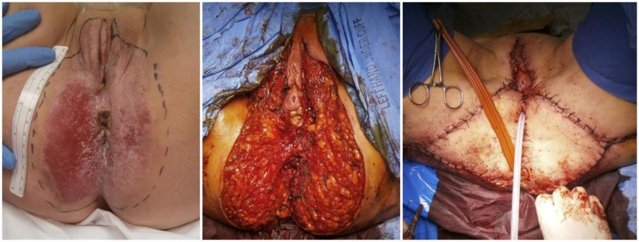
A 58-year-old woman presented with a 12-year history of misdiagnosed intra-epithelial vulvar Paget’s disease, without co-morbidities. Her medical history had begun with vulvar itching that also involved the anal region. She tried various treatments, both topical and systemic (steroids, hyaluronic acid, tocopherol, anti-fungal topical treatment) for misdiagnosed vulvar lichen sclerosus (Table 1). In October 2014, biopsy was performed and it was consistent with vulvar Paget’s disease. The patient presented to us with a large erythematous area of about 8–9 cm, spreading from the posterior hemi-vulva to the anus, with an irregular surface and whitish areas. At anoscopy, the lesion was found to extend into the anus for approximately 1 cm. In February 2015, the patient underwent laser vaporization of the anal and perianal mucosa and a simple vulvectomy with skin grafts, with two large triangular flaps bilaterally (V-Y plastic) (Figure 3). Lymphadenectomy was not performed since locoregional lymph nodes were clinically and radiologically negative. The definitive pathological report confirmed a massive localization of intra-epithelial Paget’s disease, multiple micro-infiltrations and a focus of lymph vascular space invasion. In February and October 2016, the patient presented with two locoregional recurrences, managed with a skinny vulvectomy and a topical therapy with imiquimod, respectively. In December 2018, the vulvoscopy showed an asymmetrical eczematoid area involving all the skin from the fork to the anus, towards the buttock posteriorly and laterally to the right and left, about 5×6 cm wide; biopsy revealed intra-epithelial Paget’s disease with HER2 positivity. Because of the recurrence and only partial response to imiquimod, a third surgery with a permanent colostomy was proposed, but the patient declined.

Case 4. Pre-surgery workup (left); after excision (center); and at the end of surgery after reconstruction (right).
In order to avoid bowel diversion, considering the HER2 positivity, treatment with trastuzumab was offered. In February 2019 the patient started 3-weekly trastuzumab (Figure 4A). Initially, due to no evidence of invasive components in the previous two biopsies, chemotherapy with paclitaxel was omitted. During treatment, she reported a reduction of the itching in the perianal area, with objective local response at the physical examination. In August 2019, a biopsy confirmed the persistence of intra-epithelial Paget’s disease with microfoci of stromal infiltration. Despite this initial clinical benefit, in November 2019 the locoregional situation worsened (Figure 4B), so she started a combination treatment of paclitaxel–trastuzumab. The patient experienced improvement in symptoms’ control and objective local response. Treatment was well tolerated, and no other adverse effects have been observed until the last administration. Considering the subjective relief in symptoms, maintenance therapy with 3-weekly trastuzumab was started in early May 2020 (Figure 4C).

(A,B,C) Case 4. Disease spread before the start of trastuzumab (A); at progression during trastuzumab treatment (B); and after 22 weekly paclitaxel–trastuzumab administrations (C).
DISCUSSION
Rare tumors can have disease-specific oncogenic drivers that are also present in other more common cancers for which targeted treatments already exist. This is the case for the sub-population of patients with HER2-positive vulvar Paget’s disease, who could benefit from targeted anti-HER2 therapy. In the largest series yet published, 25% cases of vulvar Paget’s disease showed an HER2 amplification,3 whereas in Paget’s disease of the breast, HER2 molecular sub-type seems to be dominant.8 This report describes three cases of metastatic and one case of recurrent HER2-positive vulvar Paget’s disease effectively treated with weekly paclitaxel and trastuzumab. In addition to case reports of patients with HER2 positive, non-vulvar extramammary Paget’s disease treated with anti-HER2 agents,9–14 there are four separate case reports of women with metastatic HER2 positive vulvar Paget’s disease treated similarly (online supplemental table). Our series, therefore, represents the largest number of reported cases of vulvar Paget’s treated with these agents.
The patients presented here had long disease courses before the trastuzumab-based therapy and had previously been exposed to multiple extensive surgeries and topical treatments. All four patients experienced objective and subjective benefits from the anti-HER2 therapy. In particular, case 1 with lung metastases had a complete radiological response at first-line therapy with a progression-free survival of about 36 months. Moreover, she experienced a partial response after a re-challenge with paclitaxel and trastuzumab with a benefit of 14 months. This patient is currently receiving treatment with trastuzumab emtansine. Case 2 started the targeted therapy after a rapid disease progression during cisplatin treatment, and had a partial response on the large pelvic mass with a benefit of about 10 months. Case 3 is, to our knowledge, the first case of metastatic vulvar Paget’s disease with central nervous system involvement. She developed intracranial progression during maintenance with trastuzumab, with stable disease in other sites. As for patients with advanced HER2 positive breast cancer in intracranial progression and systemic stability during anti-HER2 treatment, 3-weekly trastuzumab was continued and radiotherapy was performed. Finally, case 4 is emblematic because trastuzumab single agent was initially proposed as a rescue strategy to avoid colostomy after two biopsies showing intra-epithelial vulvar Paget’s disease, but despite symptom relief in the first few months, the clinical benefit was modest, lasting approximately 6 months. When paclitaxel was added to trastuzumab the objective response on the locoregional lesion was greater, as was the clinical benefit; these observations suggest a synergism between trastuzumab and taxanes, as has been established in other diseases including breast cancer.15
The failure of single-agent trastuzumab in advanced vulvar Paget’s disease was reported by Hanawa et al5 and should be taken into account when offering anti-HER2 therapy. The present experience in using paclitaxel–trastuzumab in HER2 positive vulvar Paget’s disease re-inforces the need for a tailored clinical trial aimed to assess unequivocally the effectiveness of this therapy. Unfortunately, a clinical trial of trastuzumab in this setting has been withdrawn, probably for difficulties in accrual (NCT01427244). Finally, although intriguing, the role of pertuzumab in HER2 positive vulvar Paget’s disease is still elusive since no case report using this antibody has yet been described.
CONCLUSION
This is the largest series of HER2-positive advanced vulvar Paget’s disease that, despite the small number of patients, supports the value of a combined approach with weekly paclitaxel–trastuzumab. Moreover, the aggressive nature of the HER2 positive sub-group is corroborated by the capacity to metastasize to the central nervous system. Accordingly, some conclusions can be drawn. First, testing the HER2 expression at vulvar Paget’s disease diagnosis seems to be crucial since it can influence the surgical approach and follow-up of patients due to the greater aggressiveness of the HER2 positive disease. Second, targeted anti-HER2 therapy with weekly paclitaxel and trastuzumab should be considered the first option in metastatic vulvar Paget’s disease or when other treatments for invasive recurrence are precluded. The strong biologic rationale for using anti-HER2 treatment, instead of other agents, is now supported by several case reports. Finally, future studies should investigate possibility of using anti-HER2 therapy as an adjuvant strategy to reduce the risk of recurrence after surgery.
Acknowledgments
We thank Valerie Matarese for editing the manuscript.
References
Footnotes
Twitter @Barto_Med
FS and FP contributed equally.
Contributors MB conceived the study. MB, RM, MDS, LB, and MGV wrote case reports. FS supervised the surgical aspects of the work. FP supervised the medical aspects of the work. All authors contributed to data interpretation, wrote, revised, and approved the final version of the manuscript.
Funding The authors have not declared a specific grant for this research from any funding agency in the public, commercial or not-for-profit sectors.
Competing interests FP reports grants from AstraZeneca, grants, personal fees and other from Roche, personal fees and other from Eli Lilly, personal fees from Amgen, personal fees from Ipsen, personal fees from MSD, personal fees from Takeda, grants and other from Eisai, other from Novartis and Pfizer, outside the submitted work; the other authors have nothing to disclose.
Patient consent for publication Not required.
Provenance and peer review Not commissioned; externally peer reviewed.
Data availability statement All data relevant to the study are included in the article or uploaded as supplementary information. All data are included in the main article.