Article Text

Abstract
Introduction/Background Ewing’s sarcoma/primitive neuroectodermal tumor (ES/PNET) family of tumours are rare group of malignant neoplasms. Commonly, they arise from skeletal system, but also they can arise from extraskeletal tissues.
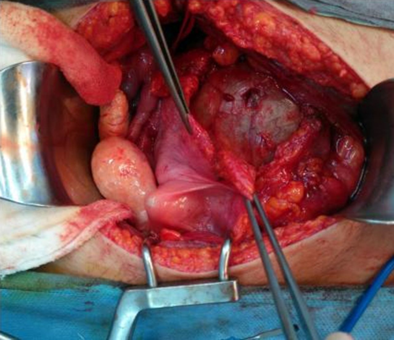
Methodology A 19-year-old nulliparous female admitted to our outpatient clinic with low abdominal pain. Abdominal ultrasound and MRI findings were revealed an adnexial solid-cyctic mass mimmicking an ovarian neoplasm. After a thorough diagnostic laparatomy; a 14 cm mass arising in the Retzius space with normal internal genitalia were observed. The tumour was firmly adhered to the bladder, urethra and other adjacent tissues. Total resection of the tumour was performed. The frozen section revealed small round blue cell tumour. The patient discharged in a good conditon on the post operative fifth day and she underwent adjuvant chemotheraphy with VAC/IE (vincristine, doxorubicin, cyclophosphamide alternating with ifosfamide, and etoposide). Immunohistochemical analysis revealed negative staining for the CD45, pancytokeratin, chromogranin, CD56, WT1, desmin, myogenin, myoD1, and diffuse positive staining for the CD99. Fluorescence in situ hybridization confirmed EWSR1 gene translocation in the 81.9% of tumour cells. Based on these histopathological, IHC and FISH findings, an extraskeletal Ewing’s sarcoma was diagnosed.
Results Here, we report a case of Ewing sarcoma of the Retzius space treated by surgery and adjuvant chemotheraphy. To the best of our knowledge, this extremely rare clinical condition is the first reported case in English literature.
Conclusion Extraskeletal Ewing’s sarcoma in the Retzius space is extremely rare. It should be included in the differential diagnosis if frozen section reveals small round blue cell tumours. Immunohistochemical analysis and FISH could help to diagnose this rare clinical situation.
Disclosure Nothing to disclose.

Ewing MR

Op photo