Article Text

Abstract
Introduction/Background Uterine adenosarcoma is a rare and aggressive malignancy. It is defined as a biphasic tumor composed of both benign glandular component and malignant stromal component. Here we report early recurrence of a uterine adenosarcom.
Methodology A Case Report.
Results A 46-year-old woman presented with vaginal bleeding and admitted to the outpatient-clinic. Her past medical history was noncontributory. Bimanual examination revealed a normal uterus and bilateral adnexa. Pelvic color ultrasound showed that a 57 × 38 mm heterogeneous mass occupying the uterine cavity. Fractional endometrial curettage revealed no malignancy. Total abdominal hysterectomy was performed. The diagnosis of the frozen section was uterine adenosarcoma. The patient subsequently received bilateral salpingo-oophorectomy, pelvic and para-aortic lymphadenectomy. Peritoneal cytology revealed nonmalignant cells. Histopathological final diagnosis was adenosarcoma with heterologous elements (cartilage and rhabdomyoblasts). Largest tumor diameter was 6 cm, lymphovascular space invasion was positive, and the tumor was in the inner half of myometrium. There was no metastasis in ovaries and lymph nodes. According to the International Federation of Gynecology and Obstetrics (FIGO) (2009) staging system, the final stage was Ib. She was discharged without postoperative treatment.
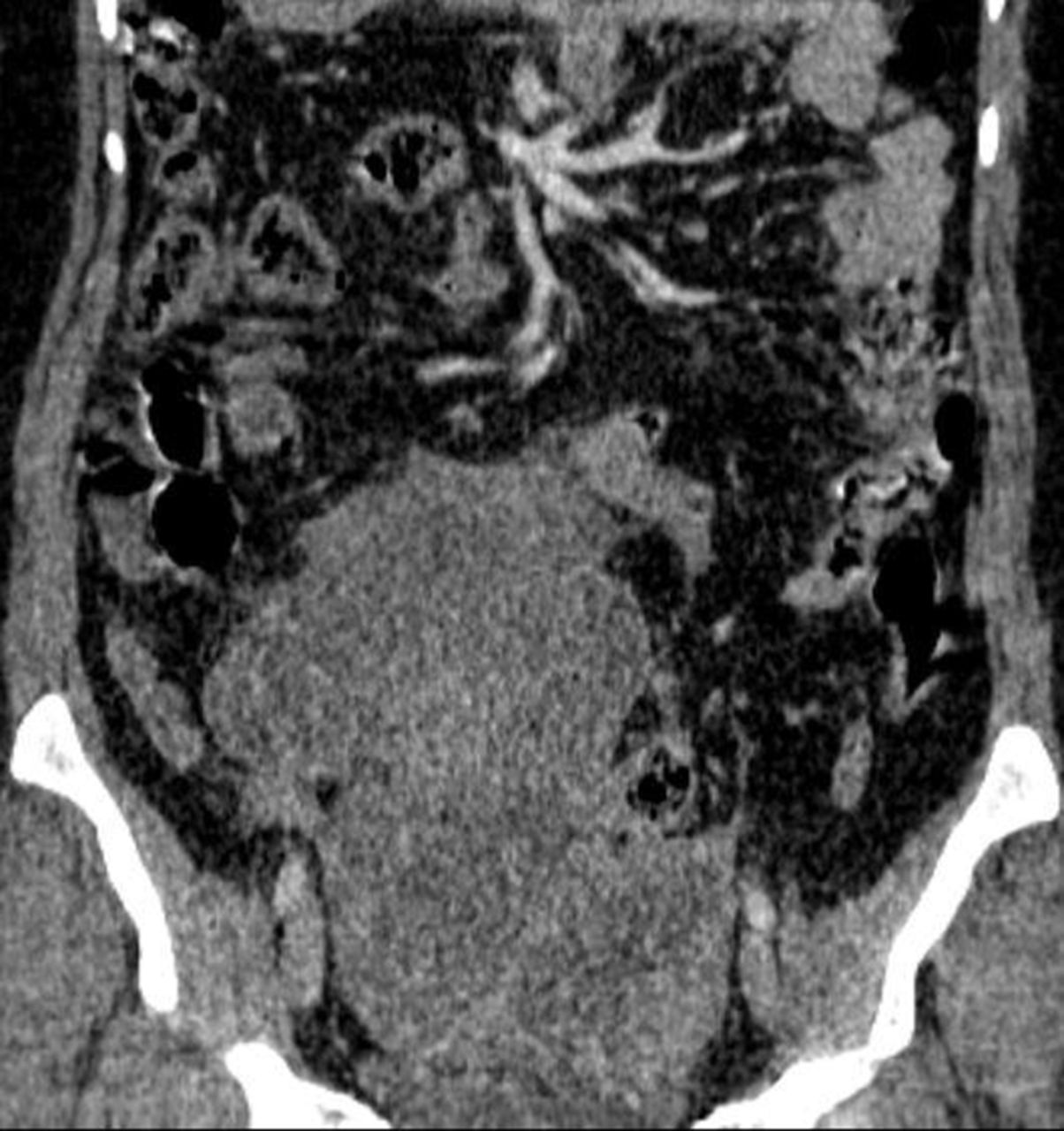
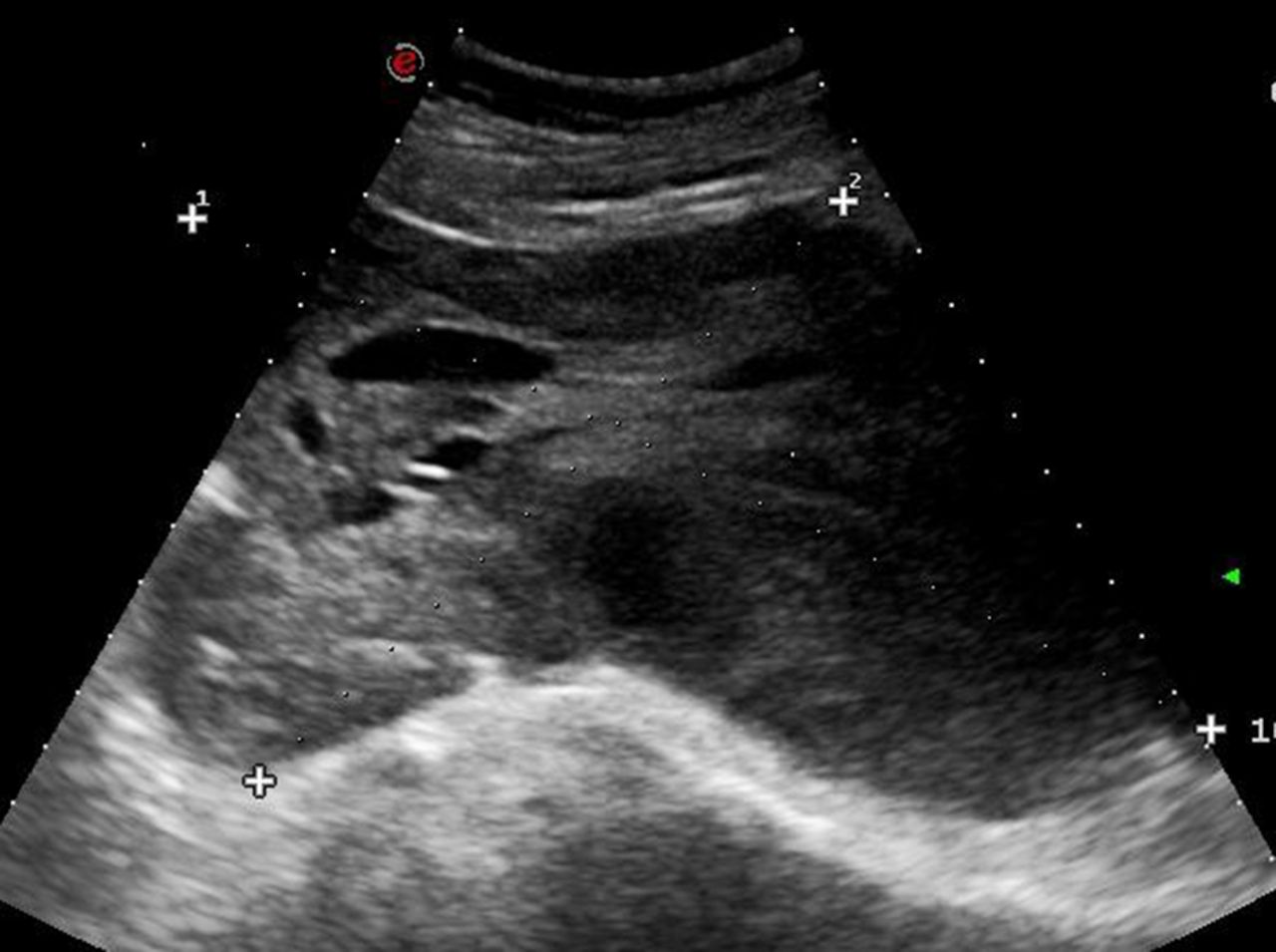
A few days before first follow-up visit, the patient was admitted again for abdominal pain. Abdominal ultrasound scans revealed an enlarged pelvic mass with a 17 × 10 cm diameter (figure 1). Contrast-enhanced Computed Tomography imaging of the pelvis revealed a large pelvic mass with hypodense areas that indicate cystic componentand peritumoral fat stranding (figure 2). The tumor was filling the pelvic cavity (18 width × 15 depth × 12 length cm) and extends into the abdominal cavity. A second surgery was planned but the patient refused to undergo surgery.
Conclusion Close follow-up of the uterine adenosarcoma patients is recommended because of the high chance of early recurrence.
Disclosure Nothing to disclose.

Longitudinal transabdominal US scan demonstrates heterogeneous hypoechoic solid mass in the pelvis
Computed Tomography imaging of the pelvis revealed a large pelvic mass with hypodense areas