Article Text

Abstract
Introduction/Background Adult granulosa cell tumours (aGCT) constitute a rare subtype of ovarian cancer, with >90% of tumours characterized by the FOXL2 c.402C>G mutation. Recurrences occur in nearly 50% of patients and are associated with a poor prognosis. Surgery is the mainstay of treatment, since the effect of adjuvant therapies are limited. Chemotherapy and hormone therapy response is difficult to predict and patient numbers insufficient to conduct informative clinical trials. Patient-specific aGCT organoid and 2D culture establishment could evaluate the effect of novel therapeutic options on a relatively large scale and enable personalized treatment in this neglected patient group.
Methodology Samples from 46 tumours (4 primary and 42 recurrences) from 18 patients were collected, mechanically homogenized to single cells and small tissue pieces, and seeded in 50% Cultrex Basement Membrane Extract on day of surgery. Basal medium (DMEM/F12) supplemented with various growth factor combinations promoted organoid formation. Medium was refreshed every 3–4 days and cells passaged every 3 weeks. In parallel, 2D monolayer cell cultures were established to perform drug screens for later validation in the more physiologically relevant organoids as they became available. Organoid and 2D tumour origin verification is ongoing by FOXL2 c.402C>G PCR.
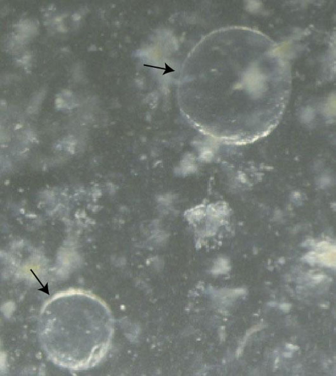
Results Tumour tissue cultivation resulted in 3D structures that remained viable for 3–4 passages, with an establishment rate of approximately 75% in primary and 26% in recurrences. Morphology of masses ranged from grape-like to cystic, with primary organoids demonstrating a more cystic morphology (figure 1) that could be robustly passaged. Monolayer cultures in DMEM/F12/FBS were successfully established for future drug screening with carboplatin, paclitaxel, tamoxifen and other first-line treatments.
Conclusion Patient-derived aGCT organoids can be established. Organoid establishment is more successful for primary tumours than for metastases, and organoid establishment optimization is necessary. Tumour cell culture establishment and drug screens are likely to provide insight into patient-specific treatment response.
Disclosure Nothing to disclose.

Organoids (black arrows) derived from primary tumor tissue. Passage III