Article Text

Abstract
Introduction The solute carrier family 12 member 5 (SLC12A5) gene is playing a putative oncogenic role in colorectal carcinoma. However, the status of SLC12A5 amplification and expression in ovarian carcinoma and its potential clinical and/or prognostic significance has not yet been investigated.
Methods In the present study, semi-quantitative staining and fluorescence in situ hybridization were used to investigate SLC12A5 protein expression and gene amplification levels. Samples were obtained from archival, formalin-fixed, paraffin-embedded pathological specimens consisting of 30 normal ovaries, 30 ovarian cystadenomas, 30 borderline ovarian tumors, and 147 invasive ovarian carcinomas. SLC12A5 immunohistochemical staining results, pathological parameters, and patient prognosis were then evaluated using various statistical models. Patient survival rate was also assessed using receiver-operator curve analysis.
Results Our results revealed no SLC12A5 protein overexpression in normal ovaries. However, 7% of cystadenomas had SLC12A5 protein overexpression along with 17% of borderline tumors and 37% of ovarian carcinomas (P<0.01). Amplification of SLC12A5 was detected in 10.3% of ovarian carcinomas. Further correlational analyses showed that SLC12A5 protein overexpression in ovarian carcinomas was significantly associated with ascending histological grade, pT/pN/pM status, as well as FIGO stage (P<0.05). A subsequent univariate survival analysis of our ovarian carcinoma cohorts resulted in a significant association between SLC12A5 protein overexpression and decreased patient survival (44.3 and 85.9 months for high and low SLC12A5 protein expression, respectively; P<0.001). Importantly, additional multivariate analysis revealed that SLC12A5 protein expression was a significant, independent prognostic factor for overall survival in ovarian carcinoma patients (P=0.003).
Conclusions Collectively, these findings support the conclusion that SLC12A5 protein overexpression could indicate an invasive and/or aggressive phenotype of ovarian carcinoma. Future work will need to investigate whether SLC12A5 protein can serve as an independent prognostic molecular marker in patients with ovarian carcinoma.
- ovarian carcinoma
- SLC12A5
- prognosis
This is an open access article distributed in accordance with the Creative Commons Attribution 4.0 Unported (CC BY 4.0) license, which permits others to copy, redistribute, remix, transform and build upon this work for any purpose, provided the original work is properly cited, a link to the licence is given, and indication of whether changes were made. See: https://creativecommons.org/licenses/by/4.0/.
Statistics from Altmetric.com
HIGHLIGHTS
SLC12A5 protein is overexpressed in primary ovarian carcinoma.
Overexpression of SLC12A5 may play an important role in carcinogenesis of ovarian carcinoma.
SLC12A5 may serve as a novel adverse prognostic biomarker for patients with primary ovarian carcinoma.
Introduction
Ovarian cancer is the most lethal and the second most frequently invasive gynecological malignancy worldwide. Recently, reports showed that an estimated 238 700 new cases and 151 900 deaths from ovarian cancer occurred in the world each year.1 2 The majority (approximately 70%) of ovarian cancer patients are diagnosed at an advanced stage of the disease (FIGO III/IV stage) due to no effective screening tests or the appearance early symptoms.3 Respectively, the 5-year overall survival rate is approximately 30%, while the rate for patients diagnosed at early stage (stage I/II) is almost 90%.1 3 Therefore, there is an urgent need to find specific and sensitive biomarkers for early detection of ovarian cancer.
Solute carrier family 12 member 5 (SLC12A5), also known as potassium chloride cotransporter 2 (KCC2), is crucial to the maintenance of neuronal chloride homeostasis.4 SLC12A5 protein overexpression is significantly associated with more advanced tumor stages.5–7 The SLC12A5 gene was amplified in human colorectal carcinoma. Other work showed that in colorectal carcinoma, DNA amplification is one of the leading causes of SLC12A5 protein overexpression. SLC12A5 upregulation promotes tumorigenesis and metastasis, thereby suggesting that SLC12A5 could serve as a new independent prognostic factor for patients with colorectal carcinoma.5 8 The role of SLC12A5 in the development and progression of ovarian cancer remains unclear.
In this study, we mainly investigated the expression of SLC12A5 protein and amplification status of the SLC12A5 gene in ovarian cancer. The clinical and prognostic significance of SLC12A5 protein expression were then analyzed using various statistical models. Our results may provide useful clinical data in the treatment of ovarian cancers.
Methods
Human tissue specimens
A total of 237 human ovarian tissue samples were taken on surgical resection in the Cancer Center and the First Affiliated Hospital, Sun Yat-sen University, Guangzhou, China. These samples included 30 ovarian tissues, 30 cystadenoma, 30 borderline, and 147 ovarian cancers tissues. All the samples were made into paraffin-embedded tissue specimens after normal dehydration and dehydration processing. All patients had not received any preoperative chemo-radiation, and patients had complete clinical follow-up data available for review. Additional clinico-pathological characteristics are summarized in Table 1. We obtained written, informed consent from all patients as well as approval from the Institute Research Medical Ethics Committee of Sun Yat-sen University prior to beginning the study.
Association of SLC12A5 expression with patient’s clinico-pathological features in ovarian carcinomas
Construction of tissue microarray and immunohistochemistry
The tissue microarray was constructed with 0.6 mm cores selected from the most representative tumor areas using a manual tissue array instrument (Beecher Instruments, Silver Spring, MD, USA), according to previously described methods.9 10 Immunohistochemical analysis of SLC12A5 was performed using a standard streptavidin-biotin-peroxidase complex method that has been previously described.5 Briefly, tissue sections were deparaffinized and rehydrated. The endogenous peroxidase activity was blocked with 3% H2O2 for 10 min. Tissue was then subjected to antigen retrieval, during which slides were immersed in 10 mM citrate buffer (pH 8.0) and boiled for 3 min in a pressure cooker. Non-specific binding was blocked by 5% normal goat serum for 10 min. The slides were incubated with a 1:100 dilution of polyclonal antibody against SLC12A5 (Abcam) at 4°C overnight in a moist chamber. Slides were then sequentially incubated with biotinylated goat anti-mouse IgG (1:100 dilution; Santa Cruz Biotechnology) and then streptavidin-peroxidase conjugate, each for 30 min at room temperature. Isotope-matched human IgG was used in each case as a negative control. Finally, the 3, 5-diaminobenzidine (DAB) Substrate Kit (Dako) was used for color development followed by Mayer hematoxylin to provide a counterstain.
To evaluate SLC12A5 staining, its extent was scored by assigning a staining percentage to positive tumor cells (0, none; 1,<20% of positive staining cells; 2, 20–50% of positive staining cells; 3,>50% of positive staining cells). Samples with scores of 0, 1, or 2 were defined as low expression, while cases with a score of 3 were defined as having high expression,5 All histological evaluations were carried out in a double-blind manner by two expert pathologists (C-JW and DX).
Fluorescence in situ hybridization
Two-color fluorescence in situ hybridization (FISH) was used to study the genetic amplification of SLC12A5. A Spectrum BAC clone (RP11-465L10) containing the SLC12A5 gene was labeled with Spectrum Red (Vysis, Downers Grove, IL). Simultaneously, a chromosome 20 centromere probe labeled with Spectrum Green (Vysis, Downers Grove, IL) was used as an internal control. The FISH reaction was carried out as previously described with slight modifications. Briefly, deparaffinized ovarian carcinoma tissue sections were treated with proteinase K (400 µg/mL) at 37°C for 45 min, followed by denaturation in 70% formamide, 2×SSC at 75°C for 8 min. Fifty nanograms of each probe were mixed in a 20 µl hybridization mixture (containing 55% formamide, 2×SSC, and 2 µg human Cot1 DNA), denatured at 75°C for 6 min, and then hybridized to the denatured tissue sections at 37°C for 24 hours. Slides were counterstained with 1 µg/mL DAPI in an anti-fade solution and were examined with a Zeiss Axiophot microscope equipped with a triple-band pass filter. FISH signals from a minimum of 300 tumor cells were evaluated per specimen. SLC12A5 gene amplification in tumor cells was defined as either: the presence of six (or more) SLC12A5 gene signals; or more than three times as many gene signals than seen in the reference signals (Figure 1D).

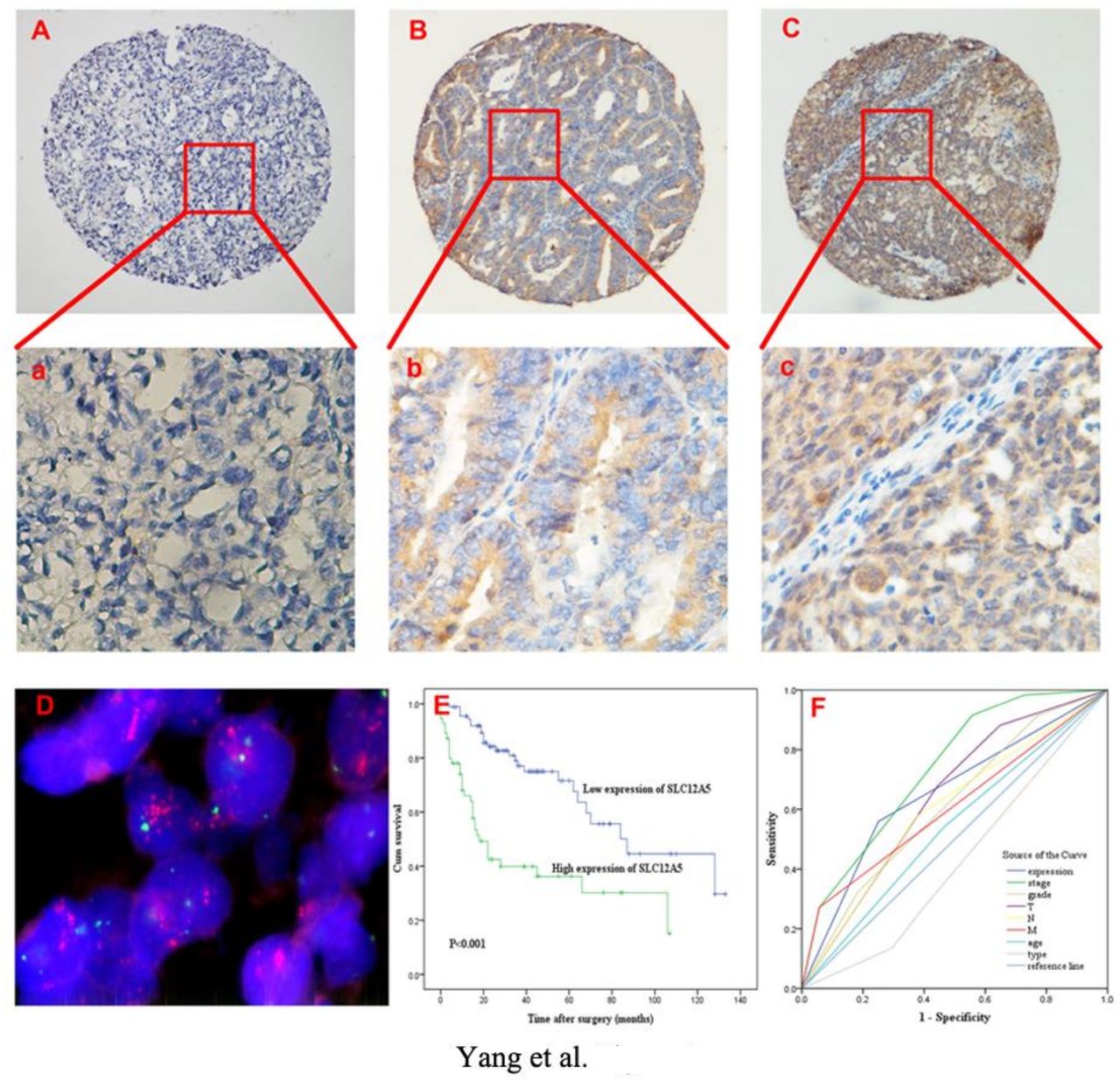
Immunohistochemical staining of SLC12A5 protein and FISH assay of SLC12A5 gene in human ovarian carcinoma tissue. (A) An ovarian carcinoma (case 24) showed normal expression of SLC12A5 protein with a negative staining of SLC12A5 protein. (B) Representative image of primary ovarian carcinoma (case 11) showing low SLC12A5 protein expression. Less than 10% of tumor cells showed high SLC12A5 nuclear immunoreactivity (original magnification,×100). (C) Representative image of SLC12A5 protein overexpression in primary ovarian carcinoma (case 145). All of the tumor cells revealed positive SLC12A5 immunostaining (original magnification,×100). (a), (b), and (c) are higher magnifications of the indicated areas from the representative images presented in (A), (B), and (C), respectively (original magnification,×400). (D) Amplification of the SLC12A5 gene was observed using FISH in the same ovarian carcinoma case (case 145). SLC12A5 gene amplification (red) was at least three times stronger than the signals for chromosome 20 centromere (green) (original magnification,×1000). (E) Probability of patient survival: low expression of SLC12A5, n=92; overexpression of SLC12A5, n=55 (P<0.001). (F) FIGO stage (AUC)=0.737,P<0.001), SLC12A5 protein expression ((AUC)=0.655, P=0.002), pT ((AUC)=0.641, P=0.004), pN ((AUC)=0.601, P=0.039), pM ((AUC)=0.607, P=0.028), Pathological grading ((AUC)=0.612, P=0.021) was significantly associated with survival.
Statistical analyses
All statistical analyses were executed using SPSS software (SPSS Standard version 20.0, SPSS Inc.). A chi-square test was used to assess the association of SLC12A5 expression with clinico-pathological features of ovarian carcinoma patients. The Kaplan–Meier method was used for univariate survival analysis and a log-rank test was used to compare different survival curves. Multivariate Cox regression analysis was used to assess the overall survival power of these parameters. Receiver operating characteristic (ROC) curve analysis were used to assess differences between the clinico-pathological features and estimation of survival predictions. The P value of<0.05 was considered statistically significant.
Results
SLC12A5 protein expression in ovarian cancers
According to the result of immunohistochemistry SLC12A5 protein was located in both the nucleus and cytoplasm of ovarian epithelial cells and ovarian cancer cells. Positive SLC12A5 expression was observed in 55 out of 147 in invasive carcinomas, whereas positive SLC12A5 expression was detected in five out of 30 borderline tumors, and two out of 30 cystadenomas (Figure 1A–C). SLC12A5 protein expression was significantly higher in invasive carcinomas than borderline tumors and cystadenomas (P<0.01, Table 2).
The expression of SLC12A5 in normal ovaries and in benign and malignant epithelial ovarian tumors*
Association of SLC12A5 protein expression with ovarian carcinoma clinico-pathological features
SLC12A5 protein expression was significantly correlated with histological grade, pT/pN/pM status, and FIGO stage (P<0.05, Table 1). There was no significant correlation between SLC12A5 protein expression or other clinico-pathological features, such as the patient age or tumor histological type (P>0.05, Table 1).
Relationship between clinico-pathological variables, SLC12A5 protein expression, and survival
Survival factors were taken into account to confirm the representativeness of the ovarian carcinomas. Several clinico-pathological parameters including tumor histological grade (P=0.002), pT/pN/pM status (P<0.01), and FIGO stage (P<0.001) revealed significant impact on patient prognosis (Table 3). The mean survival time for patients with high SLC12A5 protein expression was 44.3 months, while it was 85.9 months for those patients with low SLC12A5 expression (P<0.001, Table 3, Figure 1E).
Clinical pathological parameters and expression of SLC12A5 for prognosis of 147 patients with ovarian carcinoma by univariate survival analysis (log-rank test)
Independent prognostic factors of ovarian carcinoma: multivariate COX regression analysis
We next confirmed whether the variables observed to have prognostic impact in our univariate analysis were covariates. The correlation between the protein expression of SLC12A5 and clinico-pathological parameters (tumor histological grade, pT/pN/pM status, and FIGO stage) were assessed in a multivariate analysis (Table 4). A Cox regression model indicated that SLC12A5 expression (relative risk: 2.265, CI: 1.321 to 3.882, P=0.003), pN status (P=0.018), and FIGO stage (P<0.001) were independent prognostic factors for adverse overall survival (Table 4).
Multivariate analysis on overall survival (COX regression model)
Relationship between clinico-pathological variables, SLC12A5 protein expression, and ovarian carcinoma patient survival
To maximize the specificity and sensitivity of the results, a ROC for each clinico-pathological parameter displayed a point on the curve closest to (0.0, 1.0). The ROC analysis for each clinico-pathological variable and SLC12A5 protein expression (area under the curve (AUC)=0.655, P=0.002) was then conducted to assess patient survival status (Figure 1F).
Amplification ofSLC12A5 in ovarian tumortissue microarray
FISH analysis was informative in 87/147 (59.2%) of ovarian carcinomas, 16/30 of borderline ovarian tumors, and 14/30 of ovarian cystadenomas. Those samples that had no FISH signal, that displayed weak target signals, or that had strong background expression were categorized as non-informative cases. FISH results revealed that SLC12A5 amplification was detected in none of the ovarian cystadenomas and borderline tumor tissues. However, it was detected in 10.3% (9/87) of the informative ovarian carcinoma cases. SLC12A5 protein overexpression was observed in each of the nine cases with SLC12A5 amplification (Figure 1D). Among the remaining 78 informative cancers without SLC12A5 amplification, 50 (64.1%) cases displayed low protein expression of SLC12A5, while the other 28 (35.9%) cases showed SLC12A5 protein overexpression.
Discussion
The SLC12A5 gene, also known as potassium chloride cotransporter 2, is a gene located at 20q13.12. The amplification of 20q is one of the most frequent chromosomal aberrations in human cancers, including breast, ovarian, and gastric cancers.11 12 Recent studies show that it is frequently amplified in colorectal carcinoma, leads to tumorigenesis and metastasis in several kinds of cancer.4 5 8 However, the expression status of SLC12A5 in ovarian carcinomas has yet to be investigated and its clinico-pathological and/or prognostic values in ovarian carcinomas remain unclear. In our study, we mainly investigated whether the SLC12A5 gene was related to tumorigenesis and metastasis in epithelial ovarian cancer. Our results suggested the SLC12A5 gene and protein expression were important in the clinical characterization and prognosis of these patients.
The results showed that SLC12A5 protein was expressed higher in tumor specimens than normal specimens, suggesting that SLC12A5 protein may be involved in tumorigenesis. SLC12A5 protein expression is significantly correlated with histological grade and clinical stage (pT/pN/pM and FIGO stage), but not correlated with age or tumor histological type. These findings indicated that SLC12A5 protein is not only involved in the progress of epithelial ovarian cancer, but that it may also be a useful prognostic marker.
The abnormal SLC12A5 protein expression at both gene and protein levels is significantly associated with poor prognostic indicators and clinical outcome in colorectal cancer.5 However, there is no data on the prognostic status and clinical outcome of SLC12A5 expression in ovarian cancer. The Kaplan–Meier curves and multivariate Cox proportional hazard regression analysis demonstrated SLC12A5 overexpression was a predictor for adverse overall survival in patients with ovarian carcinoma—independent of histological grade and clinical stage. These data suggested SLC12A5 upregulation as a reliable biomarker for ovarian cancer prognosis.
Gene amplification is a common pathological mechanism of oncogene overexpression in human cancers.13 With respect to the mechanism of SLC12A5 up-regulated protein expression in ovarian carcinomas, FISH was used to determine SLC12A5 gene amplification status. The results showed that among the 87 informative cases simultaneously detected by both IHC and FISH, SLC12A5 overexpression was observed in all (9/9) ovarian carcinomas that showed SLC12A5 amplification. SLC12A5 amplification was not observed in the other 28 ovarian carcinomas with SLC12A5 protein overexpression. Hence, the upregulated SLC12A5 protein expression is complicated and might be regulated by other molecular mechanisms, including transcriptional and post-translational regulation. Further research should be carried out to probe the mechanism.
Previous studies using breast cancer cell lines have shown that SLC12A5-modulated cell spreading, migration, and invasion are not dependent on the canonical role of SLC12A5 as a K+–Cl− co-transporter. It operates by manipulating focal adhesion formation by way of an ion transport-independent mechanism.6 In addition, functional in vitro and in vivo studies have shown that SLC12A5 contributed to colorectal carcinogenesis by inhibiting apoptosis via AIF-dependent and EndoG-dependent apoptotic signal pathways. Moreover, that it promoted metastasis by regulating key elements of the matrix architecture.5 Based on these data, we hypothesize that the effect of up-regulated SLC12A5 protein expression in human cancers is complicated and likely regulates metastasis and apoptosis through several pathways. However, the potential mechanisms of how SLC12A5 functions in ovarian carcinoma have not been identified. Clearly, further studies are needed to precisely elucidate the mechanisms by which SLC12A5 promotes the development and progression of ovarian carcinomas.
In summary, this study evaluated the protein expression levels and amplification patterns of SLC12A5 in a large series of human ovary samples, including normal tissue, benign adenomas, borderline tumors, and malignant epithelial ovarian tumors. We demonstrated that SLC12A5 overexpression may be critical in the acquisition of a more aggressive biological behavior in ovarian carcinoma. As such, it has potential as a novel, reliable, and independent prognostic biomarker indicative of adverse patient outcomes.
References
Footnotes
G-PY and W-PH contributed equally.
Contributors G-PY conducted the immunohistochemical assays, evaluated the clinical records, and drafted the manuscript. W-PH assisted in conducting the immunohistochemical assays and drafting of the manuscript. J-FT, Z-XY, R-RF, N-FM, and F-WW participated in the statistical analysis as well as in its coordination. LC and YL assisted in conducting the immunohistochemical assays. Z-SY and DX aided in study design, analysis, and in data interpretation. G-FY guided the study design, evaluation of immunohistochemical results, and wrote the manuscript. All authors read and approved the final manuscript.
Funding This study was funded by grants from the Nature Science Foundation of China (No.81772769) and from the Research Project in the Science and Technology Bureau in Guangzhou (No. 201704020125).
Competing interests None declared.
Patient consent for publication Not required.
Ethics approval This study was approved by the Research Ethics Committee of the First Affiliated Hospital, Sun Yat-sen University (approval no. 2018‑46). Informed consent was obtained from each patient before study. All procedures performed in studies involving human participants were in accordance with the ethical standards of the First Affiliated Hospital, Sun Yat-sen University.
Provenance and peer review Not commissioned; externally peer reviewed.
Data availability statement All data relevant to the study are included in the article or uploaded as supplementary information.