Article Text

Abstract
Placenta accreta spectrum disorders are a major risk factor for severe postpartum hemorrhage and maternal death worldwide, with a rapidly growing incidence in recent decades due to increasing rates of cesarean section. Placenta accreta spectrum disorders represent a complex surgical challenge, with the primary concern of massive obstetrical hemorrhagic sequelae and organ damage, occurring in the context of potentially significant anatomical and physiological changes of pregnancy.
Most international obstetrical organizations have published guidelines on placenta accreta spectrum, embracing the creation of regionalized ‘Centers of Excellence’ in the diagnosis and management of placenta accreta spectrum, which includes a dedicated multidisciplinary surgical team. One mandatory criterion for these Centers of Excellence is the presence of a surgeon experienced in complex pelvic surgeries. Indeed, many institutions in the United States and worldwide rely on gynecologic oncologists in the surgical management of placenta accreta spectrum due to their experience and skills in complex pelvic surgery.
Surgical management of placenta accreta spectrum frequently includes challenging pelvic dissection in regions with distortion of anatomy alongside large aberrant neovascularization. With a goal of definitive management through cesarean hysterectomy, surgeons require a systematic and thoughtful approach to promote prevention of urologic injuries, embrace measures to secure challenging hemostasis and, in selected cases, employ conservative management where indicated or desired.
In this review recommendations are made for gynecologic oncologists regarding the management and important considerations in the successful care of placenta accreta spectrum disorders. Where required, gynecologic oncologists are encouraged to be proactively involved in the management of placenta accreta spectrum, not only intra-operatively, but also in the development of clinical protocols, guidelines, and pre-operative counseling of patients, as a ‘call if needed’ approach is suboptimal for this potentially major and life-threatening condition.
- Gynecologic Surgical Procedures
- Surgical Procedures, Operative
Statistics from Altmetric.com
Introduction
Placenta accreta spectrum disorders have evolved into one of the major iatrogenic public health challenges of the 21st century. In the United States (US), more recent rates have quadrupled since the 1980s, reaching a high of approximately 1 in 272 deliveries in 2016.1
The genesis is based on an increasing rate of uterine surgeries, predominantly prior cesarean section. The incidence increases with the number of prior cesarean sections ranging from 0.3% in pregnancies with one previous cesarean section, increasing to up to 6.7% for those with five or more cesarean sections.2 Furthermore, when placenta previa is also present, the risk of placenta accreta spectrum is approximately 11% with one prior cesarean section, 40% with two prior cesarean sections, and as high as 60% with three or more prior cesarean sections.3
Overall, maternal morbidity from placenta accreta spectrum has been reported to range from 24% to 67% and includes massive hemorrhage (average estimated blood loss ranging from 2000 mL to 4000 mL) with associated risks of large volume blood transfusion, coagulopathy, visceral injury, infection, thromboembolism, and need for reoperation.4 5 Reports on the incidence of maternal mortality range from 0.05% to as high as 7%, influenced by factors including location and resources, antenatal diagnosis, and dedicated multidisciplinary management by an experienced team.6 7
Most national and international obstetrical organizations including the International Federation of Gynecology and Obstetrics (FIGO), the American College of Obstetricians and Gynecologists (ACOG), the Society for Maternal-Fetal Medicine (SMFM), and the Society of Obstetricians and Gynecologists of Canada (SOGC), have published placenta accreta spectrum guidelines embracing an experienced multidisciplinary protocol-based team approach through the creation of regionalized ‘Centers of Excellence’ in diagnosis and management.8–10 One of the essential criteria of a Center of Excellence is the presence of a surgeon experienced in complex pelvic surgeries, commonly a gynecologic oncologist.8–10
Competent skills and expertise in pelvic surgery is a core requirement for the management of placenta accreta spectrum disorders, particularly in safe retroperitoneal dissection, ureterolysis, pelvic artery ligation (usually uterine or internal iliac artery), cystoscopy, ureteral stent placement, and meticulous uterovesical plane dissection particularly in cases with parametrial involvement.11
It should be noted that advanced pelvic surgeons in obstetrics and gynecology, other than gynecologic oncologists, with the appropriate skills and training may also perform the primary surgical role in many centers. In fact, the specific credentials of the surgeon (gynecologic oncologist, general obstetrician-gynecologist, maternal-fetal medicine specialist) are likely less important than the criteria of consistent, ongoing experience with cases of placenta accreta spectrum.12 However, as gynecologic oncologists are frequently called on to provide this service, in both high- and low-resource settings, it is important to provide an evidence-based review of optimal intrapartum management to facilitate and guide the gynecologic oncology community to play a substantive role, where indicated.
Role of the Gynecologic Oncologist
While the primary role of a gynecologic oncologist is the comprehensive management of individuals with gynecologic cancers, their surgical skills have historically been utilized for both emergency and complex obstetrical surgeries.
In a tertiary-level Australian study, the authors reported on non-gynecologic oncology surgeries which required the presence of a gynecologic oncologist, where obstetric indications represented the vast majority. The main specific indications within this group were placental abnormalities, followed by massive obstetrical hemorrhage.13
Furthermore, the recently published ACOG/SMFM placenta accreta spectrum guideline was also endorsed by the Society of Gynecologic Oncology (SGO), highlighting the importance of the involvement of gynecologic oncologists in the management of this condition.9
Recently, Hoffman et al reviewed the past, present, and future of surgical training requirements in gynecologic oncology fellowships. The authors identified the operative management of placenta accreta spectrum as a priority area of competence for graduating fellows given the increasing frequency and complexity of such cases.14 Indeed, many institutions in the US and worldwide rely on gynecologic oncologists in the surgical management of these disorders.
Highlighting this, the five institutions in the University of California Fetal Consortium have instituted a multidisciplinary approach to placenta accreta spectrum: four institutions have a mix of maternal-fetal medicine, obstetrician, and gynecologic oncologist surgeons on their placenta accreta spectrum teams. Only one institution relied on maternal-fetal medicine and obstetric surgeons, calling gynecologic oncology only if required intra-operatively.
In a retrospective chart review of pathologically confirmed cases of placenta accreta spectrum from 2009 to 2014 in the University of California Fetal Consortium including 151 confirmed cases, gynecologic oncology surgeons were involved in 58% of surgeries (n=71). This involvement resulted in a statistically significant lower estimated blood loss when adjusting for final pathology (2.2 L vs 2.25 L; p=0.02).15
Another Australian study aimed to assess the role of gynecologic oncologist involvement in the management of placenta accreta spectrum by studying 98 cases of histologically confirmed abnormally invasive placenta and comparing outcomes of three distinct groups: (1) those who had a gynecologic oncologist present at the start of the procedure (group 1; n=43), (2) those who had a gynecologic oncologist called in during the procedure (group 2; n=23), and (3) those who had no gynecologic oncologist involved (group 3; n=32). The median estimated blood loss for the entire cohort was 2150 mL (range 300–11 500 mL). Group 2 had a significantly higher blood loss than the other groups (median 4400 mL; p=0.001) . Transfusion requirements were higher in groups 2 and 3 compared with group 1 (p=0.004). Other maternal and neonatal morbidity was similar across all three groups. The authors concluded that the early presence of a gynecologic oncologist at delivery when placenta accreta spectrum is suspected is to be promoted and that a ‘call if needed’ approach is not acceptable for these complex cases.16
In an Egyptian study focusing on the conservative management of placenta accreta spectrum, the authors found that the involvement of a gynecologic oncologist in the surgical procedure led to lower maternal morbidity (17.5% vs 72.2%; p<0.001) with less estimated blood loss, less packed red blood cell units, and less operative time. Furthermore, the rate of successful uterine preservation was higher when a gynecologic oncologist was present (100% vs 50%; p<0.001).17
Finally, beyond involvement in surgical management, in some institutions gynecologic oncologists are key members in developing clinical protocols, quality improvement initiatives, and pre-operative clinical management for each case of suspected placenta accreta spectrum. In these centers, patients with a high suspicion for placenta accreta spectrum are referred to a gynecologic oncologist for comprehensive antenatal consultation and surgical planning.18
FIGO Classification of Placenta Accreta Spectrum Disorders
Placenta accreta is defined as a spectrum of abnormal placentation disorders. Depending on the depth of uterine trophoblast invasion, three subtypes have been pathologically differentiated: (1) superficial placenta accreta, where there is decidual (endometrial) deficiency and the anchoring placental villi attach directly onto the myometrium without deeply invading it; (2) placenta increta, where the villi penetrate deeply into a myometrial defect with partial myometrial thinning or dehiscence; and (3) placenta percreta, where the villi reach or extend beyond the uterine serosa with complete loss of overlying myometrium and possible invasion into surrounding structures or organs.19 It should be noted that different subtypes of placenta accreta usually co-exist in the same specimen and that an area can be focal or diffuse.
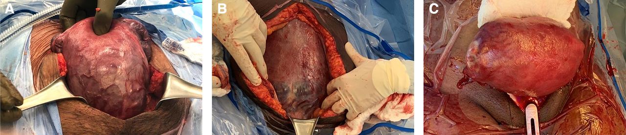
To support the process of clarifying reported data on placenta accreta spectrum in the international literature, in 2019 FIGO developed a clinical grading system (Table 1).20 Most importantly, this grading system refers to a classification and not a staging system, to differentiate it from cancer terminology within the gynecologic oncology context. Examples of surgical findings are demonstrated in Figure 1A–C.
International Federation of Gynecology and Obstetrics (FIGO) general classification of placenta accreta spectrum disorders

(A) A 20-week gestation pregnancy at laparotomy demonstrating anterior placenta previa increta (International Federation of Gynecology and Obstetrics (FIGO) grade 2 placenta accreta spectrum). Note the lower segment distension with bluish/purple coloring and increased neovascularity. Placenta increta confirmed histopathologically. (B) A 35-week gestation pregnancy at laparotomy demonstrating anterior placenta previa percreta (FIGO grade 3c placenta accreta spectrum). Note the lower segment distension with bluish/purple coloring, placental tissue seen at the uterine serosa along with increased neovascularity. Placental villi were noted to be invading the broad ligaments bilaterally. Placenta percreta confirmed histopathologically. (C) A 38-week gestation pregnancy post-cesarean at laparotomy demonstrating right cornual placenta percreta (FIGO grade 3a placenta accreta spectrum). Note the right cornual distension with bluish/purple coloring, Placental tissue seen at the uterine serosa along with increased neovascularity. Placenta percreta confirmed histopathologically.
Multidisciplinary Management and Pre-Operative Planning
Dedicated multidisciplinary team-based care in the diagnosis and management of placenta accreta spectrum disorders to improve outcomes is now recognized as the standard of care. There will, however, be some cases in which diagnoses are missed or are emergently managed with or without the presence of an experienced team. In such cases, outcomes have been shown to be poorer, highlighting the importance of optimizing diagnosis, pre-operative planning, and multidisciplinary care.21
The ideal composition of these multidisciplinary teams may consider consultation by the following core specialties: obstetrics, maternal-fetal medicine, surgical gynecology or gynecologic oncology, anesthesia, neonatology, and perinatal mental health services.22 Additional teams which may also require consultation include hematology, urology, general surgery, vascular surgery, and intensive care.
Accurate diagnosis is an essential step to achieving improved outcomes. The mainstay of diagnosis involves adequate screening of at-risk individuals, with subsequent specialized ultrasound imaging using both transabdominal and transvaginal approaches. Overall, the diagnostic performance of ultrasound for placenta accreta spectrum disorders is excellent, with a sensitivity of 90.72% (95% CI 87.2 to 93.6) and specificity of 96.94% (95% CI 96.3 to 97.5).23 In some cases, particularly those with posterior, fundal, or parametrial involvement, non-contrast magnetic resonance imaging (MRI) may be of added utility, with overall reported sensitivity of 94.4% (95% CI 86.0 to 97.9) and specificity of 84.0% (95% CI 76.0% to 89.8%).24
The role of pre-operative and proactive patient blood management strategies is essential in all obstetric cases where surgical blood loss is expected to be high. Iron stores and hemoglobin levels should be assessed at diagnosis and optimized, along with screening for hemoglobinopathies and red cell antibodies. Hematology consultation may be required in such cases where indicated. Our group, and others, also advocate the use of intra-operative cell salvage where available, to reduce the need for allogenic blood transfusion.22
Antenatal anesthesia consultation is paramount, providing expert care in the prevention and management of massive obstetrical hemorrhage. Historically, general anesthesia was utilized in most cases. In recent times, however, contemporary groups are successfully utilizing regional anesthesia epidurals with or without combined spinal anesthesia for the duration of cesarean hysterectomy for placenta accreta spectrum.8 Regional compared with general anesthesia, where appropriate, appears to be associated with decreased hemorrhage-related morbidity, blood transfusion requirements, and neonatal respiratory complications.23 Patient satisfaction may also be improved if they are conscious for the birth experience of their infant.
Optimizing timing of delivery remains a challenging aspect of care, balancing the increasing risk of unplanned emergent delivery at term with the perinatal risks of iatrogenic prematurity if delivered pre-term. Most recently, the International Society for Abnormally Invasive Placenta (IS-PAS) suggested expectant management until after 36 weeks’ gestation for asymptomatic patients with no obvious risk factors for pre-term delivery, whereas planned delivery at around 34 weeks’ gestation was advised for those with a history of previous pre-term birth, recurrent vaginal bleeding, short cervix, or pre-term pre-labour rupture of membranes.25
Non-Conservative Surgical Management (Cesarean Hysterectomy)
Cesarean section followed sequentially by immediate hysterectomy without attempting removal of the placenta (placenta left in situ) is considered the gold standard for definitive treatment of placenta accreta spectrum.
Type of Incision
The ideal surgical incision for a planned cesarean hysterectomy allows for sufficient access to the uterus, provides optimal exposure to surrounding structures, and avoids the placenta to reduce potential blood loss. A midline skin incision is recommended by some groups, as this facilitates a high fundal uterine incision, which can avoid the upper margin of anterior placentas which extend superiorly.8
A low transverse skin incision may be adequate in situations where the superior margin of the placenta does not enter the upper segment of the uterus, or if uterine conservation is planned, but may not provide sufficient exposure in more severe cases. Other options for abdominal entry include the Joel-Cohen incision, Cherney extended transverse incision, and a modified Maylard (high transverse) incision.26
Techniques for Hysterectomy
Total abdominal cesarean hysterectomy with salpingectomy is the recommended surgical method for emergent peripartum hysterectomy, and the preferred option in the management of placenta increta or percreta if cervical involvement is present. This approach is also typically required if there is significant lower uterine segment or cervical bleeding (Table 2).
Subtotal abdominal cesarean hysterectomy with salpingectomy is another common approach and has been reported to result in shorter operating time, decreased blood loss, reduction in blood transfusions, and lower peri-operative complications.8 However, as this approach leaves a cervical stump, there remains a potential risk of cervical dysplasia and malignancy, the need for regular cervical cytology, and other associated issues such as bleeding, vaginal discharge, or infection. In addition, there has been no evidence that subtotal hysterectomy provides protection against urinary tract injury when compared with total hysterectomy in surgeries specifically for placenta accreta spectrum disorders.27
Five key steps of cesarean hysterectomy for placenta accreta spectrum disorders*
Internationally, Centers of Excellence have developed additional novel techniques for cesarean hysterectomy to minimize hemorrhage and reduce unintentional urinary tract injury (Table 3). The gynecologic oncologist A E Selman proposed a posterior retrograde hysterectomy via the pouch of Douglas, which, he suggests, allows for safe resection of the involved urinary bladder in cases of placenta percreta with intraluminal bladder involvement.28 Shamshirsaz et al described a modified radical hysterectomy (without oophorectomy) technique that includes extensive use of a bipolar cautery device to assist with early surgical devascularization.29 Their retrospective study compared this approach with a non-standardized approach for severe placenta accreta spectrum, reporting lower median estimated blood loss (2.1 L vs 3.5 L, respectively; p=0.031), with a trend to fewer blood transfusions ≥4 units (60% vs 87%, respectively; p=0.056), and were less likely to be delivered emergently (23% vs 64%, respectively; p=0.001). The use of disposable devices such as a linear cutting autostapler,30 a vessel sealing device,31 or a combination of both32 have also been suggested as innovative approaches to reduce blood loss and rates of blood transfusion; however, outcomes have not yet been reported comparing these various techniques.
Planned delayed hysterectomy is an alternative approach to the cesarean hysterectomy. This method may be utilized in circumstances of severe placenta percreta where extensive invasion of surrounding structures would impact the ability to safely perform an immediate hysterectomy. Delayed hysterectomy, which is typically performed between 3 and 12 weeks postpartum,33 may facilitate placental reabsorption, decrease vascularity, and use the normal process of uterine involution which may simplify future surgery, allow the use of minimally invasive surgical approaches, and reduce overall surgical morbidity. However, this approach comes with an associated risk of coagulopathy, hemorrhage, and sepsis in the interim period. Zuckerwise et al compared outcomes between delayed hysterectomy in 14 patients (five before the scheduled date due to complications, 36%) versus 20 patients who underwent immediate cesarean hysterectomy (four due to significant peri-operative bleeding, 20%).18 The median (IQR) sum-total estimated blood loss for delivery and delayed hysterectomy was significantly lower than the immediate hysterectomy group (1.3 L (0.1–2.2 L) vs 3.0 L (2.4–4.3 L), respectively; p<0.01). Delayed hysterectomy was associated with fewer patients requiring transfusion of ≥4 units of blood than the immediate cesarean hysterectomy group (14.2% vs 45%, respectively; p=0.016). There was one maternal death in each group (incidence 7% for delayed hysterectomy and 5% for immediate hysterectomy).
Novel techniques for cesarean hysterectomy in patients with placenta accreta spectrum disorders
Conservative Management
In certain situations, uterine conservation may be successfully achieved, either leaving the placenta in situ (termed expectant management) or with placental removal combined with partial uterine resection and repair. Potential candidates for uterine conservation include those in three main groups.10 First, those who strongly prefer to retain their fertility and are adequately counseled regarding the potential risks and chance of successful future pregnancy. The second group who may be amenable to uterine preservation include those with extremely severe disease on imaging, where there is evidence of extra-uterine invasion into adjacent structures including the bladder lumen, bowel, or broad/deep parametrium. This population is at increased risk of significant surgical complications at the time of delivery including massive hemorrhage and organ damage. Third, there may be infrequent cases where there is very focal disease detected or placenta accreta spectrum located in the upper uterine segment which may be amenable to wedge resection and approximation of vital myometrium to achieve hemostasis.
Uterine Conservation Techniques
To achieve uterine conservation with expectant management, following delivery of the infant, the umbilical cord is resected, suture ligated proximal to the placenta, and the uterus closed in a routine fashion. Whether or not to use prophylactic or therapeutic uterotonics, uterine compression sutures, uterine artery ligation, or intrauterine balloon tamponade remains largely debated in the literature.16 34 Delayed surgical options are also reported with limited evidence. Following delivery, delayed hysteroscopic resection of remaining placental tissue has been utilized to definitively treat the condition or reduce complications of retained placenta.35 Another approach, mentioned previously, involves delayed hysterectomy; however, this method is not routinely recommended or supported by most expert consensus groups.
There are two clinical contexts in which uterine conservation with placental removal or placenta accreta spectrum resection may be safely achievable. The first is in cases of focal disease with a clearly delineated area of placenta accreta spectrum (usually percreta with myometrial dehiscence) bordered by viable myometrium without disease. With placental removal and resection up to the viable myometrium, the uterus can then be oversewed or revised with healthy tissue.36 Similarly, the Triple-P procedure has also been proposed to have favorable success with pelvic devascularization, followed by placental removal with en-bloc uterine revision and then suturing of viable tissue.37 The second clinical context is in cases of fundal or posterior placenta accreta spectrum. Obstetric hemorrhage from these placental bed sites is more amenable to conservative surgical approaches, medical management, or embolization techniques to successfully control bleeding. If a hysterectomy is still required, the procedure can usually be performed with relative ease compared with anterior placenta accreta spectrum disorders.
Complications with Conservative Management
The analysis of outcomes from cases managed expectantly are challenging to interpret from the predominantly retrospective literature as due to the lack of histopathological confirmation, false-positive diagnoses are likely to be included in their analyses. This may potentially lead to more favorable reported outcomes and provide a source of bias. A systematic review of 44 studies reporting outcomes with expectant management of 72 patients found that severe complications were common, developing in 40 (56%), with uterine preservation achievable in 42 (58%) patients38. Neither prophylactic uterine artery embolization nor the use of chemotherapy (methotrexate) improved success rates, with one reported case of maternal death directly related to methotrexate use. The mean time for complete placental resorption was lower in the embolization group than in the non-embolization group (22.4 weeks vs 35.3 weeks; p=0.014). Either emergency or elective hysterectomy was performed at a mean of 44.6 days after cesarean delivery; and among the 23 patients with hysterectomy-related complications, 18 (78%) experienced bladder injury, intra-operative bleeding (>2.0 L), or both.
With a potentially prolonged course of recovery, frequent follow-up, and requirements to reside in close proximity to emergent care, expectant management can pose significant challenges. Among the potential adverse outcomes reported during the time of placental reabsorption and resolution, severe vaginal bleeding (53%), sepsis (6%), and death (up to 4%) are the most significant risks.38 Sentilhes et al conducted a retrospective study into fertility and pregnancy outcomes following conservative treatment.39 They reported subsequent amenorrhoea due to severe intrauterine synechiae (Asherman syndrome) in 8.3%, with a further 5.2% describing oligomenorrhoea. Of the 27 (28%) participants who wanted more children, 24 (89%) had 34 pregnancies (21 third-trimester deliveries, one ectopic pregnancy, two terminations, and 10 miscarriages) with a mean time to conception of 17.3 months (range 2–48 months). All 21 deliveries resulted in healthy babies born after 34 weeks of gestation, with placenta accreta spectrum recurring in 6/21 cases (29%).
Measures to Minimize Surgical Blood Loss
Multidisciplinary use of measures aimed at minimizing blood loss, which include percutaneous vessel balloon placement, transfusion medicine interventions, and surgical and pharmacological measures, are essential in the surgical management of placenta accreta spectrum disorders.40 The routine implementation of protocol-based individualized care has led to significantly reduced maternal hemorrhagic morbidity and should be considered the standard of care.10 Blood conservation measures include administration of peri-operative intravenous tranexamic acid (anti-fibrinolytic) at skin incision, and its use has been demonstrated to reduce intra-operative blood loss.41 Intra-operatively, the return of blood collected via cell salvage during surgery is recommended when possible and available.
Meticulous surgical dissection at each of the surgery’s key steps is paramount to minimizing blood loss. Strategic ultrasound-guided hysterotomy to deliver the fetus, with selective use of an auto-stapler device or efficient single-layer closure may also reduce surgical blood loss at the time of delivery. Hemorrhage during superior devascularization of the uterus is often minimal with standard suture ligation; the principal risk of bleeding comes from excessive upward traction on the uterus by lateral straight clamps rather than by manual elevation.
Dissection of the uterovesical plane is often a key step where significant surgical bleeding from large aberrant neovascularization is first encountered; a meticulous lateral-to-medial dissection of this plane is required. Division of these engorged blood vessels and adipose layer down with the bladder is the preferred approach. Careful dissection should be continued inferiorly until the level of the anterior vaginal fornix where colpotomy can be performed. Securing hemostasis of the vaginal vault, followed by prompt vault closure, is often the final surgical step associated with significant blood loss.
Exposure of the parametrial and pelvic side wall anatomy prior to bladder dissection, a critical surgical step that allows identification of the ureters, has repopularized the strategy of routine or selective ligation of the anterior divisions of the internal iliac arteries with an associated reduction in blood loss.11 Recently, members of our group reported that internal iliac artery ligation, as part of a multidisciplinary protocol-based quality improvement initiative, can achieve very low median blood loss (1.0 L) in cases of cesarean hysterectomy for severe placenta accreta spectrum.24
Percutaneous placement of inflatable balloons within the pelvic arteries, most commonly in the anterior divisions of the internal iliac arteries, was a previously popular surgical blood loss reducing measure in many centers.42 This strategy comes at the expense of increased costs and resources, prolonging surgical care, and with potential risks of vascular injury to blood supply to the lower limbs.43 Tow randomized controlled trials have been conducted investigating the role of internal iliac artery percutaneous balloons versus control in the surgical management of placenta accreta spectrum. The first, by Salim et al, reported no significant differences between the intervention and control groups in the mean number of packed red blood cell units transfused, 5.2 (±6.2) and 4.1 (±3.8), respectively (p=0.90), or in surgical blood loss, 4950 (±5051) and 4709 (±3434) mL (p=0.72), respectively.44 The second trial, by Chen et al, also reported no difference in the number of packed red blood cell units transfused (5.3±5.3 in the intervention group vs 4.7±5.4 in the control group; p=0.54). Importantly, hospitalization costs and incidence of post-operative fever were significantly higher in the intervention group.45 With evolving surgical expertise aimed at vascular dissection and ligation of these structures as stated above, the benefit of this strategy has come into question. 44
Complex aberrant placental blood supply arising from vascular branches additional to the internal iliac arteries is a management challenge in even the most experienced surgical hands. Infrarenal aortic compression or clamping methods are often last-resort life-saving strategies that have been utilized by many teams if required.46 To address this risk, some centers have adopted an approach of routinely utilizing an infrarenal aortic balloon with short intermittent inflation, with low rates of surgical blood loss and complications.47 Because of the favorable outcomes in contemporary Centers of Excellence specializing in placenta accreta spectrum surgery, the routine use of this approach remains a matter of ongoing research and is limited to selected individuals where a demonstrated complex pelvic arterial blood supply is proven or suspected.
Urologic Considerations
Despite being considered a relatively rare obstetrical condition, placenta accreta spectrum disorders are a significant risk factor for urinary tract injuries, involving the bladder and/or ureter(s) in up to 48% and 18% of cases, respectively.8 The bladder is the most frequent extra-uterine organ involved in placenta percreta cases. Distortions of anatomy alongside aberrant neovascularization are commonly noted in these challenging cases, some leading to either unavoidable injuries with surgical dissection, or intentional cystotomy/partial cystectomy during planned placental resection of an affected posterior bladder wall. As such, FIGO has assigned stage 3b of placenta accreta spectrum classification to those with bladder invasion.20
Preventing urinary complications can be challenging as clinical symptoms of bladder invasion are rare, with gross hematuria only occurring in one quarter of cases where there is actual placental invasion into or through the bladder wall.48 Prenatal diagnosis of placenta accreta spectrum is commonly accomplished by ultrasound during the second and third trimester of pregnancy; however, the true performance of ultrasound in detecting bladder invasion is unclear, in fact only one study reported on the accuracy of the presence of a focal exophytic mass extending into the bladder in predicting bladder involvement with a high specificity of 100% but with a low reported sensitivity of 16.7%.49
MRI is highly accurate in detecting placental extra-uterine spread, and specifically bladder invasion: in one of the largest prospective studies, the most predictive MRI feature was the presence of a bladder vessel sign with a sensitivity of 95.5% and a specificity of 96.3%.50 Pre-operative cystoscopy is frequently utilized by several Centers of Excellence; however, the literature does not provide evidence for clear recommendations on this. In fact, one meta-analysis of 54 cases with bladder involvement found that pre-operative cystoscopy was not useful in predicting involvement.51 Conversely, the establishment of provisional antenatal diagnosis of urinary tract involvement was associated with a reduction in unintentional urinary tract injury in these cases when compared with incidental diagnosis at surgery (39% vs 63%; p=0.04).27 For these reasons, cystoscopy remains of high importance if bladder involvement is suspected on imaging.
The utilization of ureteric stenting may further aid identification of the ureter and prevent inadvertent transection or ligation at hysterectomy. Furthermore, in cases where an injury is not prevented, the presence of ureteric stents may lead to easier intra-operative recognition of the injury and facilitate prompt repair. One systematic review including 292 patients who underwent cesarean hysterectomy for placenta accreta spectrum reported that ureteric stent placement versus no placement was associated with a significant reduction in urologic complications (6% vs 33%; p<0.01).27 It should be noted that if ureteric stents are utilized, some authors recommend they should be placed in the operating room just prior to delivery with all resources prepared, as placement may precipitate uterine contractions, potentially leading to bleeding and requiring emergency or hurried surgery.52 Furthermore, this timing of ureteric stenting could avoid inflammatory reactions around the ureter making it challenging to identify.
Beyond cystoscopy and ureteral stent placement, meticulous surgical technique plays an important role in the prevention of urinary tract injuries. Challenging surgical dissection of the uterovesical plane is frequently encountered due to anatomical distortions, submillimeter-thin tissue planes, significant aberrant neovascularization, and pre-existing adhesions. An adjunct technique to minimize urologic complications involves intra-operative filling and emptying the bladder with 100–300 mL of methylene blue in normal saline. This may be helpful in identifying the superior bladder margin and aid in identifying and dividing aberrant blood vessels.25 Another technique to assist in separating the hypervascular bladder wall away from the extremely thin lower uterine segment involves a lateral-to-medial dissection approach. As this process evolves surgically, the operative team should aim to dissect both the engorged blood vessels and adipose layer down with the bladder rather than the uterus.11
Surgical teams must avoid unnecessary intra-operative bleeding throughout the hysterectomy. Major intra-operative bleeding limits both visibility and can create undue urgency around bladder dissection. As such, urologic injuries have been shown to increase when intra-operative blood loss is greater.29 One suggested approach to mitigate this is primary dissection of the bladder prior to delivery, and this has been shown to facilitate identification and creation of the uterovesical plane prior to a potential intra-operative hemorrhage.27 In rare cases of suspected intraluminal bladder invasion, some authors recommend deliberate cystotomy, identification of the percreta villous tissue, and excision of the involved bladder region rather than pursuing a difficult dissection.29 52 An alternative option in some patients with severe bladder invasion is conservative management achieved by leaving the placenta in situ and performing delayed hysterectomy or expectant management for placental reabsorption, as described previously.27 In conclusion, depending on the suspected extent and localization of urinary tract involvement, urologists play an important role in prevention and treatment of ureteric injuries and in reconstructive surgery of the bladder and ureter, which may be required in up to one-third of these cases.53
Conclusions
Most national and international obstetrical organizations have published placenta accreta spectrum guidelines endorsing an experienced multidisciplinary protocol-based team approach through the creation of regionalized ‘Centers of Excellence’ in diagnosis and management8–10 25 40 (Table 4). The specific makeup of surgical multidisciplinary teams has been well described and includes: an experienced obstetrician (often a maternal-fetal medicine specialist), an anesthesiologist with expertise in complex obstetric cases, a surgeon experienced in complex pelvic surgery (often a gynecologic oncologist), a urologist (with experience of open urologic surgery including ureteric re-implantation), an interventional radiologist, a colorectal surgeon, a vascular surgeon, and a hematologist.25 Pragmatically, this team should be available 24 hours a day, 7 days a week, with rapid access to ensure that expertise is available for emergent cases, which frequently arise.
Summary of current recommendations from selected international guidelines
The early presence of an experienced pelvic surgeon at delivery when placenta accreta spectrum is suspected results in a significant reduction in blood loss and transfusion requirements when compared with a ‘call if needed’ approach.5
Beyond participation in surgical management, gynecologic oncologists should be central in the development of clinical protocols, guidelines, and establishing standards of care in all cases of suspected placenta accreta spectrum. Ideally, patients should be referred for antenatal surgical consultation depending on the suspected clinical and radiologic organs and regions involved. Finally, it should be highlighted that the most important aspects of the surgical team are surgical experience, expertise, and standardized procedures in placenta accreta spectrum care rather than training in any specific surgical specialty.
Ethics statements
Patient consent for publication
Ethics approval
Not applicable.
References
Footnotes
Contributors OT: review concept, writing the urologic considerations section. LA: revising the article. HFM: writing the 'Non-Conservative Surgical Management (Caesarean-Hysterectomy)' section. MAM: writing the 'Measures to Minimize Surgical Blood Loss' section. SRH: writing the 'Conservative Management' section.
Funding The authors have not declared a specific grant for this research from any funding agency in the public, commercial or not-for-profit sectors.
Competing interests None declared.
Provenance and peer review Not commissioned; externally peer reviewed.