Article Text

Statistics from Altmetric.com
Case presentation
A 63 year-old postmenopausal woman presented to her family physician with a tender palpable mass in the right lower quadrant in November 2019. She noticed that this pain was reproducible with pressure on the right hip. Her medical history was significant for hypothyroidism, tubular adenomas, and sessile serrated adenomas with low-grade dysplasia found on colonoscopy. Her surgical history was significant for laparoscopic tubal ligation, right rotator cuff repair, vaginal cervical cerclage, and left breast lesion removal for benign disease. Her family history was significant for breast and ovarian cancer on her maternal side. On abdominal examination, there was fullness in the lower abdomen and the pain was reproducible on palpation over her right anterior superior iliac spine. The cervix and vagina looked grossly normal on speculum examination. On bimanual and rectovaginal examination, the pelvic mass was felt to be high in the pelvis, mobile and tender to palpation. Her serum CA 125 was 27 U/mL (normal <34.0 U/mL). She underwent a pelvic MRI arranged by her family physician.
Dr Metser
A pelvic MRI ( Figure 1A ) in February of 2020 showed a lobulated, heterogeneous T2 hyperintense soft-tissue mass (solid arrows), measuring approximately 12.2×8 x 8.5 cm (transverse x AP x sagittal), with a 5.7 cm simple appearing cyst along its cranial aspect (dotted arrow). This well-circumscribed mass was situated posterior to the uterus without an obvious connection to the uterus and seemed to arise from the right adnexa. The mass showed heterogeneous post-contrast enhancement. There was small amount of free fluid in the pelvis, as well as nodular thickening of the peritoneal reflection in the posterior cul-de-sac. There was no evidence of pelvic adenopathy. Based on the imaging, the differential diagnosis included either a pedunculated fibroid or borderline ovarian tumor. Based on the presentation, the mass was not typical of an ovarian malignancy.

Preoperative imaging. (A) Sagittal (left) and axial (middle) T2-weighted MRI showing T2 hyperintense soft-tissue mass (solid arrows) and simple cyst along cranial aspect (dotted arrow). (B) Coronal CT image (right) demonstrating right paracolic soft-tissue nodule (solid arrow) and primary mass (dotted arrow).
The preoperative CT scan ( Figure 1B ) showed trace-free fluid in the pelvis, mesenteric nodule measuring 1.0×1.0 cm in the left upper quadrant, a small omental nodule in the left paramedian infraumbilical omentum measuring 0.4 cm, and another hypodense nodule along the right paracolic gutter measuring 1.9×0.9 cm suspicious for disease deposits. There were multiple retroperitoneal lymph nodes at the level of aortic bifurcation at the upper limits of normal in size but abnormally hyperenhancing with rounded atypical morphology. The anteverted uterus was inseparable from a large midline pelvic mass in conglomerate 12.1×8.3×13.5 cm. This mass had an inferior bilobed solid component and a superior fluid attenuation component 6.2×4.9×4.9 cm. The mass seemed to be of right ovarian origin, with right ovarian vessels communicating to the mass and probable right ovarian stroma stretched around the anterior aspect. This pelvic mass extended anteriorly to contact the anterior abdominal wall. The left ovary was unremarkable. Overall, the impression was that there was small-volume peritoneal carcinomatosis and metastatic adenopathy at the level of the aortic bifurcation. There was no evidence of thoracic metastatic disease.
Dr Heredia
Based on this presentation and imaging findings, what would be in your differential diagnosis and what recommendations would you make to the patient?
Presentation of a large, mostly solid adnexal tumor, in a postmenopausal woman is often an indication for laparotomy with frozen section. In the setting of imaging studies suggesting low-volume peritoneal and retroperitoneal involvement and negative tumor markers, the differential diagnosis should include non-epithelial ovarian tumors, specifically sex cord-stromal tumors, such as Sertoli–Leydig, adult granulosa cell tumors, and thecomas. Additionally, uterine adenosarcomas arising in endometriosis implants could be included in the differential in these MRI images. Benign pathology should also be considered such as pedunculated leiomyomas or ovarian fibromas: however, given this clinical and imaging scenario, these are unlikely.
After assessment by gynecology oncology, the patient was recommended to undergo a laparotomy, total hysterectomy, and bilateral salpingo-oophorectomy. The plan was to send the ovary for frozen section and if malignant, to proceed with a staging procedure.
Dr Laframboise
Please describe details regarding the surgical procedure
In April 2020, the patient was taken to surgery and the intraoperative findings were as follow: 250 mL of ascites, 12 cm size tumor involving both ovaries and fallopian tubes, with likely origin from the right ovary, and the cul-de-sac peritoneum. She underwent total abdominal history, bilateral salpingo-oophorectomy, pelvic peritonectomy, omentectomy, excision of para-sigmoid nodule, and right para-aortic lymphadenectomy. There were several small nodules less than 1 cm in the left upper quadrant, omentum, and right paracolic gutter which were excised. A 5 cm solid nodule adjacent to the rectosigmoid in an epiploica was also excised. There were several enlarged lymph nodes over 1 cm located at the bifurcation of the aorta which were excised. There was no visible residual disease at the end of the procedure. Frozen section of the right ovary was consistent with a stromal neoplasm.
Dr Pakbaz
On macroscopic examination, the largest tumor focus involved the right ovary and measured 13 cm at its maximum dimension. It revealed a mainly solid creamy-yellow cut surface ( Figure 2 ) with foci of papillary projections, hemorrhage, and approximately 20% of cystic component. The tumor involved both ovaries, fallopian tubes, uterine serosa, and omentum, as well as right pelvic side wall, rectosigmoid, and cul-de-sac region. The two right paraaortic lymph nodes were negative for tumor.
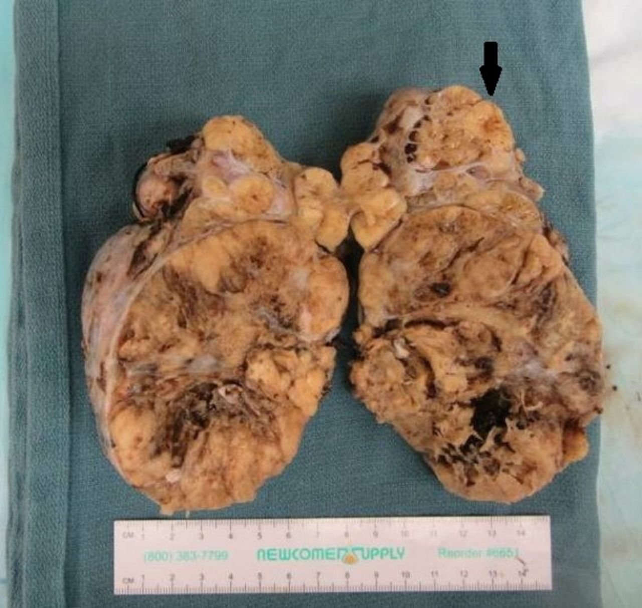
Cut surface showed a mostly solid lobulated mass containing cystic, hemorrhagic, and necrotic foci. It invaded through the ovarian surface (black arrow).
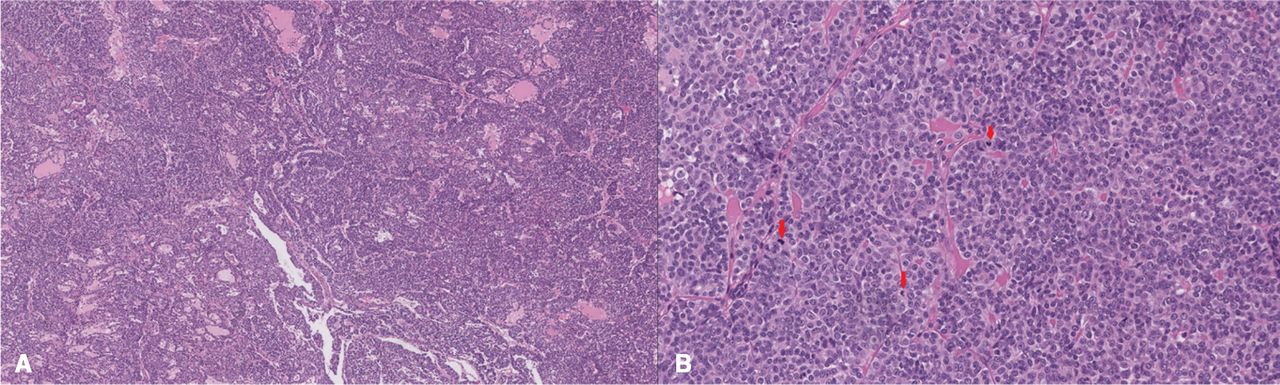
Microscopically, the tumor showed variable patterns including diffuse sheets, small closely packed hollow and solid tubules, areas with sieve-like growth pattern, and foci of necrosis ( Figure 3A ). The tumor cells were medium-sized oval to fusiform with some showing eosinophilic cytoplasm. Variable nuclear atypia, prominent nucleoli, and focal nuclear grooves were identified ( Figure 3B ). Mitotic activity was variable but in hot spots the count was up to 16/10 HPF (high-tpower field).

(A) Tumor revealed solid sheets, tubules, and sieve-like growth pattern (left). (B) Higher-power photomicrograph showing medium-sized oval tumor cells with eosinophilic cytoplasm, variable nuclear atypia, prominent nucleoli, and nuclear grooves along with increased mitotic activity (red arrows) (right).
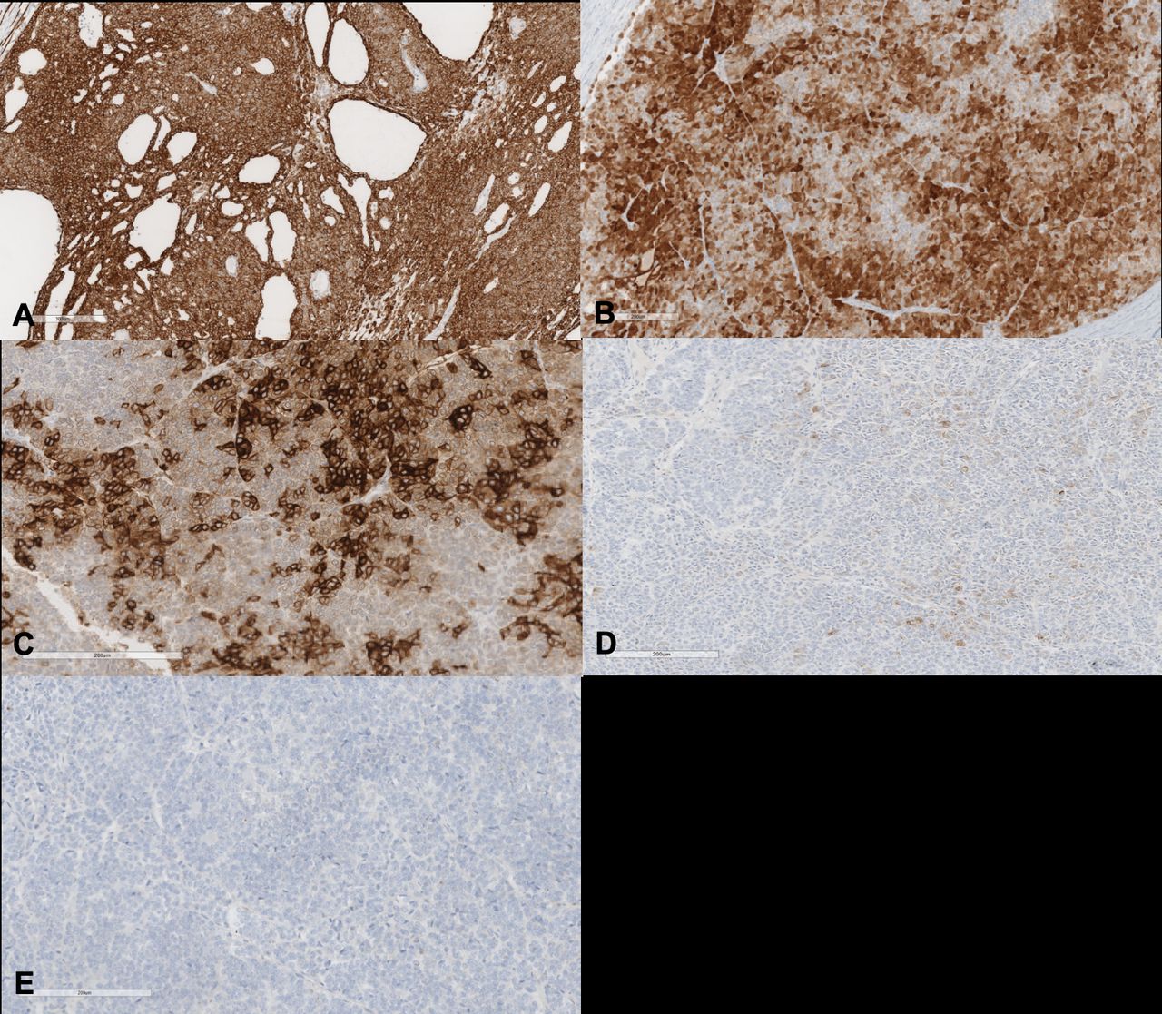
Due to morphologic overlap with more common entities in the ovaries such as sex cord stromal tumor (adult granulosa cell tumor) and ovarian surface epithelial tumor of endometrioid or serous type and possibly mesotheliomas, a comprehensive immunohistochemical panel was applied: some are illustrated in Figure 4 . Tumor cells were positive for AE1AE3, CK7 (focal), calretinin (patchy), WT-1, CD10 (focal), alpha-inhibin (scattered cells), and SF-1 (focal) and they were negative for EMA, chromogranin, synaptophysin, GATA3, TTF-1, and PAX-8. Estrogen receptor (ER) and progesterone receptor (PR) were variably expressed. The immunoexpression pattern of p53 was normal (wild type). One would usually expect more diffuse expression of alpha-inhibin in the adult granulosa cell tumor. Lack of PAX-8 and EMA immunoreactivity makes the ovarian surface epithelial tumor unlikely and the absence of squamous differentiation (within the tumor) and endometriosis and/or adenofibroma (in the background ovary) are also helpful to the decrease the possibility of endometrioid carcinoma. Negativity for EMA also argued against the diagnosis of malignant mesothelioma. In differentiating an ovarian mesonephric-like carcinoma, negative PAX8, GATA3, and TTF1 and expression of ER/PR were helpful. Overall, with this morphology and immunophenotype, the diagnosis of female adnexal tumor of probable Wolffian origin (FATWO) was rendered. Although CD117 (c-kit) was negative in the tumor, the presence of necrosis, ovarian surface invasion, increased mitotic activity, and high MIB-1 proliferation index were the worrisome features highlighting the potential for a malignant behavior in this tumor. 1
Immunohistochemical findings in tumor cells. (A) Diffuse AE1/AE3 expression (B) Patchy expression of calretinin. (C) Focal expression of CD10 (D) Scattered patchy positivity for alpha-inhibin that expected to be more diffuse in sex cord stromal tumors (E) Negativity for EMA that argued against surface epithelial tumors or mesothelioma.
After the surgery, the patient stayed for 3 days in hospital and had an unremarkable postoperative recovery.
Dr Heredia
Based on this final pathology diagnosis, what would be your recommendation for this patient?
Wolffian adnexal tumors are rare and often found incidentally on a routine pelvic ultrasound examination as a well-vascularized semisolid adnexal tumor. Usually, they present as a well-circumscribed non-adherent smooth yellow-tan unilateral mass of the broad ligament or mesosalpinx. Its size is widely variable (0.8–25 cm). If symptomatic, the main complaints are pelvic/abdominal pain, abdominal swelling, and/or a palpable mass. Symptoms of compression (urinary frequency, tenesmus, and constipation) are also reported. 2 They are typically not associated with any specific tumorous markers. No risk factors have been identified.
Final pathology reveals this tumor is a low-volume completely debulked FIGO stage IIIC female adnexal tumor of probable Wolffian origin. It is considered that complete surgical resection provides the best overall outcomes both for primary and recurrent disease.1 2
Since recurrences have been reported in up to 10%–20% of patients and the median time to recurrence is 48 months (range, 13–96), 1 3 4 I would recommend clinical examination and CT in a 4–6 months' interval or if any symptoms arise as advisable, at least for the first 2–3 years and then every year. Due to rarity of these tumors, there are no standard recommendations regarding upfront adjuvant treatment, nor salvage therapy in recurrent disease.
Closing summary
Female adnexal tumor of probable Wolffian origin was first reported in 1973 by Kariminejad and Scully in a case series including nine patients. 5 Since then, less than 100 cases have been reported. The WHO renamed them as Wolffian adnexal tumors in 2003. The Wolffian ducts, also known as mesonephric ducts, are paired embryonic structures. They develop in the male and female embryos but are only maintained in males by Sertoli cell secretion of the Antimüllerian hormone. In adult women, its remnants appear typically as asymptomatic mucosal folds or cysts in the lateral aspect of the cervix, uterine corpus, vagina, mesosalpinx, and ovarian hilium in the form of hydatids of Morgagni. Gartner’s cysts are probably the most clinically recognizable remnants of Wolff’s duct.
The age at diagnosis ranges from 18 to 87 years with a mean 50 years old.1 2 Imaging studies such as a vaginal pelvic ultrasound identify a mainly solid and well vascularized tumor. Further studies include CT which can accurately determine its origin since they can grow to reach the upper abdomen, as well as to rule out metastatic disease. MRI imaging has been reported as depicting nonspecific features, with a difficult differential diagnosis with other solid pelvic tumors such as subserosal leiomyomas and thecomas. A Japanese study retrospectively analyzed the MRI of five cases of FATWO describing a characteristic hyposignal margin in the T2 weighing, which is also present in some benign ovarian tumors but never in ovarian carcinomas. 6
Macroscopically they are sharply demarcated and encapsulated tumors. They are often solid gray-yellow or brownish tumors with areas of hemorrhage and cystic necrosis, but also lobulated, solid-cystic, or even cystic presentations have been reported. There is also a very variable intratumor architectural histologic appearance. Usually a solid pattern (with sheets of fusiform to spindle cells), a tubular pattern (with closely packed, winding, and branching tubules), and the ‘sieve-like’. In the differential, one should also consider tumors such as Sertoli cell or adenomatoid tumors and clear cell carcinomas. 2 7 The absence of Leydig cells and endocrine symptoms, along with the presence of the characteristic ‘sieve-like’ histological pattern, usually help in the differential diagnosis.
There is no specific immunohistochemical stain for these tumors. Wolffian adnexal tumors express immunoreactivity for pancytokeratin (AE1/3 100%), CAM 5.2 (100%), CK 7 (88%), keratin 903 (17%), CK8, CK18, calretinin (91%), inhibin-A (68%), and vimentin (100%). They are weakly reactive for chromogranin, synaptofisin, and neuron-specific enolase. They are usually negative for epithelial membrane antigen (EMA), S100, actin, CD15, HBME-1, and CK20. Variable expression for estrogen, progesterone, and androgen receptors as well as CD117 (C-kit) have been reported, prompting some attempts for targeted adjuvant treatment.1 7
Standard treatment for Wolffian adnexal tumors is considered to be complete surgical resection including hysterectomy with bilateral adnexectomy and tumor debulking surgery, if necessary. Although fertility-sparing surgery is possible since most tumors are limited to one ovary at presentation, most recurrences have been reported in patients formerly treated by tumor resection alone. 3 4
Adjuvant therapy has been used after primary surgery in a few cases. Ramirez et al reported the only three patients receiving radiation therapy with one recurrence. In the same publication, three patients received platinum-based adjuvant chemotherapy with one recurrence. 2 Lee Kwon et al 8 reported a case with extratumoral spread to the fallopian tube who received three cycles of adjuvant carboplatin (AUC6) + paclitaxel 175 mg/m2 and no evidence of recurrence 12 months' later. A similar case presented by Deshimaru et al 9 received the same adjuvant scheme and progressed within the first 4 months of follow-up.
In the recurrent scenario only the carboplatin + paclitaxel combination has shown evidence of at least stabilization of the disease. Wakayama et al 4 presented a case in which initial imatinib mesylate had failed as first-line treatment for recurrence. After 10 cycles of carboplatin (AUC5) + paclitaxel 180 mg/m2, the patient had a complete response. 4 In addition to this scheme, pegylated liposomal doxorubicin (50 mg/m2 every 4 weeks) has shown some response in one case but was suspended for severe drug-induced side effects. Other drugs such as irinotecan and gemcitabine used as second- or third-line chemotherapy had no effect on tumor progression. 9
Hormonal treatment attempts with leuprorelin (22.5 mg intramuscular) concurrently with chemotherapy or medroxyprogesterone acetate (400 mg orally/day for 3 months) have not shown any beneficial effect on disease response, despite hormone receptor positivity. 2 9 In the setting of ER + tumors, tamoxifen was used in a stage IV case of Wolffian adnexal tumor with pulmonary metastasis but there is no report on tumor response. 10
Lastly, in the recurrence setting, imatinib mesylate, a tyrosine kinase inhibitor effective in the treatment of a subgroup of c-Kit protein expressing (CD117) gastrointestinal stromal tumors has been used in only three cases. Steed et al 11 reported a case in which imatinib was used as adjuvant therapy after multiple surgeries and chemotherapy. After 10 months of oral treatment, the patient was free of recurrence and continued treatment. Wakayama et al 4 reported disease stabilization with imatinib after incomplete surgical resection of recurrent disease. Progression was noted after 4 months. The third reported case has no further information about disease response. 12
Footnotes
Twitter @Sarapakbaz21
Contributors SRK contributed to conception, data acquisition, drafting, revision, and final approval of the work. FH contributed to data curation, drafting, revision, and final approval of the work. SP contributed to data curation, analysis, drafting, revision, and final approval of the work. UM and SL contributed to conception, data acquisition, project administration, drafting, revision, and final approval of the work.
Funding The authors have not declared a specific grant for this research from any funding agency in the public, commercial, or not-for-profit sectors.
Competing interests None declared.
Provenance and peer review Not commissioned; externally peer reviewed.