Article Text

Abstract
Introduction/Background It is unknown if future fertility is compromised by the administration of chemotherapy during pregnancy. The aim of this study was to identify if chemotherapy affects the maternal ovaries during pregnancy, whether these effects depend on type of chemotherapy and duration of exposure, and if pregnancy protects against chemotherapy-induced gonadotoxicity.
Methodology Pregnant 8-week-old female BL6 mice (N=115) were exposed to 6 different single chemotherapeutic agents (carboplatin, cisplatin, paclitaxel, epirubicin, doxorubicin or cyclophosphamide) or saline at gestational day (GD) 13.5. The mice were sacrificed at GD 15.5 or GD 18.5. Ovaries were assessed by histopathology and immunohistochemistry. Follicle count was determined per follicle stage and per treatment modality.
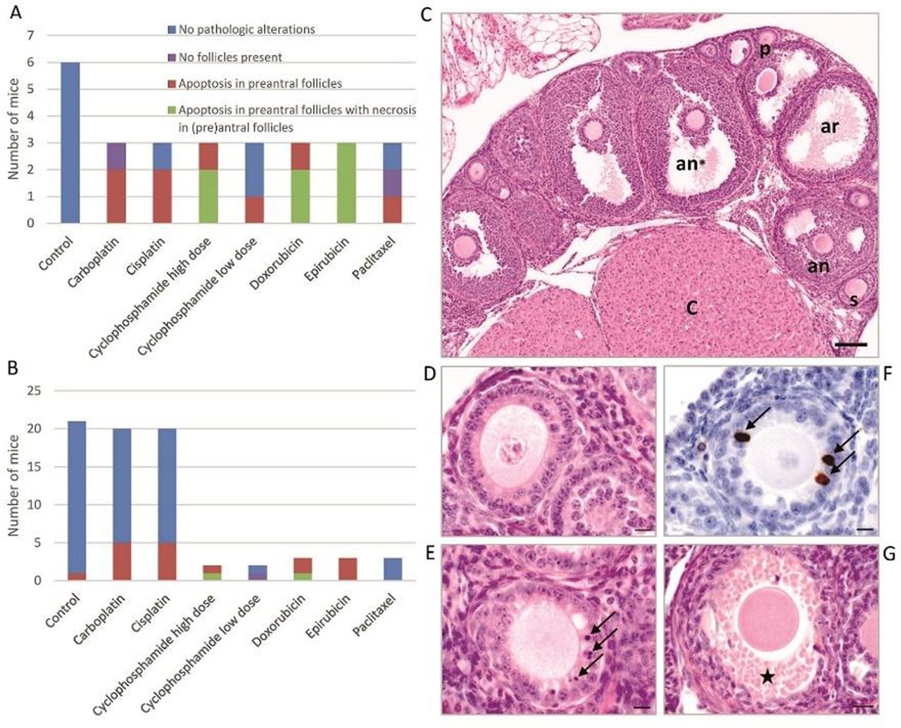
Results Maternal ovarian damage was demonstrated by the presence of apoptosis and necrosis in preantral follicles (figure 1). The extent of this damage depends on type of chemotherapy and duration of exposure (2 or 5 days). After short exposure, 81% of ovaries showed histopathologic signs of damage compared to 36% after long exposure, which might suggest a transient effect. Loss of primordial follicles (PMFs) was observed after both short and long exposure, with a reduction of more than 70%. Evidence of DNA damage, as demonstrated by phospho-H2AX expression, was present in 23% (range 0–89%) of PMFs exposed to chemotherapy, but only in the short exposure group (figure 2). Overall, the least damage was seen after administration of paclitaxel.

Histopathologic findings in ovaries of pregnant mice exposed to chemotherapy.(A) Short exposure experiment (sacrifice at GD 15.5). (B) Long exposure experiment (sacrifice at GD 18.5). (C) Overview of normal ovary containing preantral (p: primary, s: secondary), antral (an: antal, an*: antral preovulatory), and atretic (ar) follicles, and corpora lutea (c). (D) Normal preantral follicle. (E) Apoptosis in preantral follicles, arrows indicate apoptotic cells. (F) IHC of Caspase-3 in preantral follicle, arrows indicate positively stained granulosa cells. (G)Necrosis of granulosa cells of the follicle, star indicates area of necrotic cells. Scale bars: C=100 µm, D-F=10 µm, G=20 µm

Phospho-H2AX Immunohistochemistry of primordial follicles in pregnant mice of short exposure experiment. (A)) Expression versus non-expression of phospho-H2AX in primordial follicles per treatment modality (n = 6 mice in control group and n = 3 mice per chemotherapeutic agent). Examples of staining of primordial follicles in (B) control group (non-expression), (C) carboplatin group (expression), and (D) cyclophosphamide high dose group (expression). Arrows indicate primordial follicles. Scale bar: B-D = 10 µm
Conclusion Despite physiological ovarian function suppression during gestation, chemotherapy-induced damage of the ovaries occurs in pregnant mouse models, potentially affecting future fertility.