Article Text

Abstract
Introduction/Background*Systematic pelvic and paraortic lymphadenectomy is part of the initial epithelial ovarian cancer (EOC) staging surgery but this procedure is associated with potential severe morbidity. Moreover, there is no evidence suggesting a possible therapeutic value. The detection of the sentinel lymph node (SLN) in the early stage of EOC is in the experimental phase and there is little literature about it. This study evaluates the lymphatic mapping with radiotracer and indocyanine green (ICG)) and the detection of ovarian SLN.
Methodology Prospective cohort study in ovarian masses suspected of malignancy or re-staging surgery after confirmed malignancy. Albumin [99mTc] Tc-nanocolloid was injected into the utero-ovarian and infundibulo-pelvic ligaments. After 15 minutes, intraoperative images were acquired with a portable gammacamera. Subsequently, the adnexectomy and intraoperative pathological analysis were performed. In the cases in which malignancy was confirmed, ICG was injected proximally to the sectioned area of the ligaments and the lymphatic chains were traced with the fluorescence camera prior to the pelvic and paraortic lymphadenectomy. Ultrastaging of the SLN was done. The follow-up for possible adverse events lasted 30 days after surgery.
Result(s)*20 patients were included between September-2020 and May-2021. Ovarian carcinoma was confirmed in 8 (40%) cases. In all cases gamma probe was used, in 60% the gamma camera and 40% the fluorescence camera. Some SLN was detected in 18/20 cases (90%) with exclusive paraortic drainage in 5/18 (28%), pelvic in 1/18 (6%) and in both territories in 12/18 (66%). Overall para-aortic drainage was observed in 17/18 (94%) patients (35% supramesenteric, 30% at the level of the inferior mesenteric artery, and 35% inframesenteric). 100% of aortic SLNs were detected with a gamma probe after being visualized with the gamma camera. In the 8 patients who underwent lymphadenectomy, 1 case had positive nodes diagnosed by ultrastaging and the rest of the lymph nodes were negative. No complications related to the technique were observed.

Albumin [99mTc] Tc-nanocolloid injection into the utero-ovarian and infundibulo-pelvic ligaments
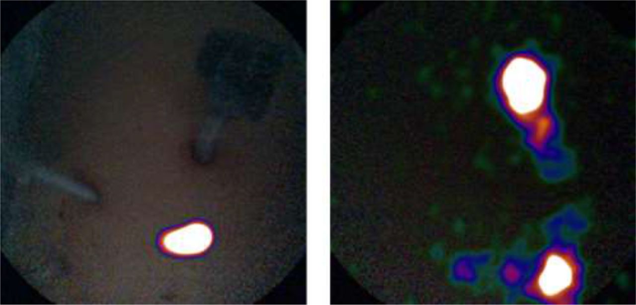
Images acquired with the portable gamma camera at 15 minutes and at 90 minutes postinjection. Paraortic drainage is observed.
Conclusion*The SLN technique is feasible and safe. The intraoperative gamma camera shows the lymphatic map and is especially useful in the paraortic region.