Article Text

Abstract
Introduction/Background*The aim of this study was to assess the diagnostic accuracy of preoperative 18F-FDG PET or PET/CT in detecting retroperitoneal lymph node (pelvic lymph node and para-aortic lymph node) metastasis in patients with high-risk endometrial cancer.
Methodology A systematic literature review was performed in PubMed and Web of science with the terms ’endometrial cancer’ and ‘positron emission tomography. Inclusion criteria were prospective or retrospective studies that evaluated lymph node metastases in the retroperitoneal area in high-risk endometrial carcinoma and provided the necessary information to construct the 2x2 table. For the quantitative analysis, the pool sensitivity and the pool specificity for node detection was calculated. The quality of the studies was assessed with the QUADAS-2.
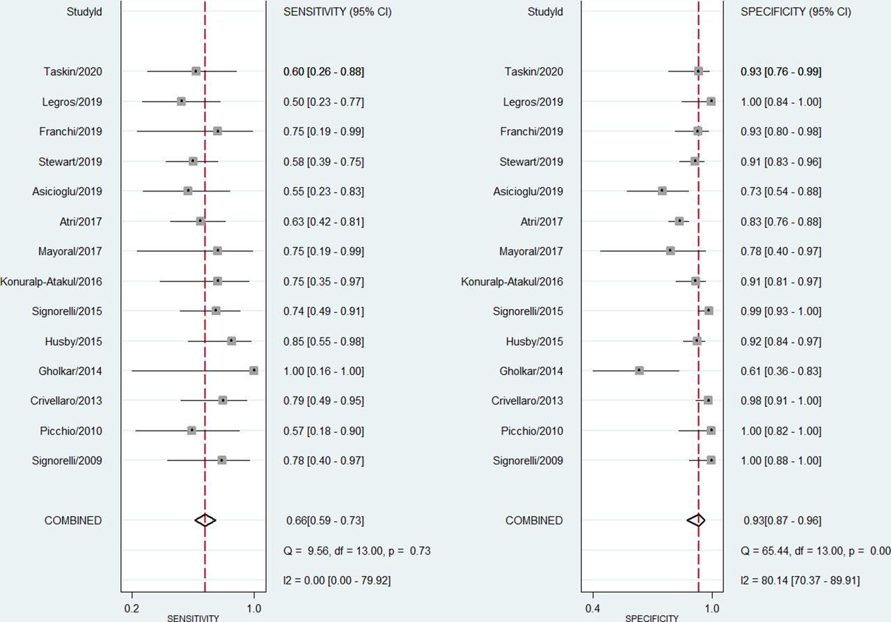
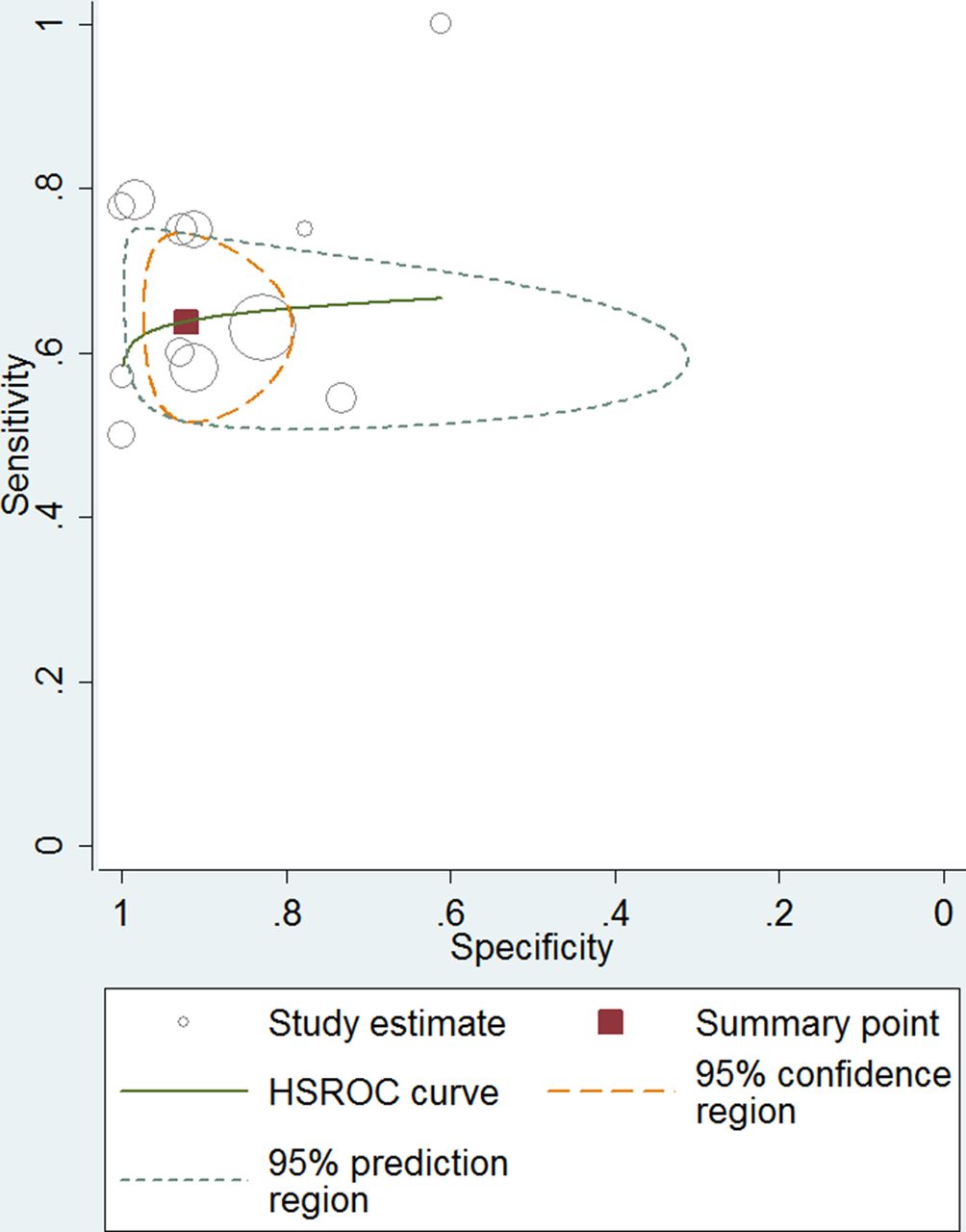
Result(s)*The research resulted in 478 eligible citations from January 1990 to May 2020. After the exclusion, fourteen articles that met all the inclusion criteria were included, comprising data from 850 patients. The sensitivity, specificity and AUC of 18F-FDG PET or PET/CT in the detection of lymph node metastases in high-risk endometrial cancer were 0.66 (95% CI: 0.59-0.73), 0.93 (95% CI: 0.87-0.96) and 0.72 respectively.

Conclusion*Despite the widespread use of 18F-FDG PET or PET/CT for imaging staging in patients with high-risk endometrial cancer, this study show the moderated sensibility of this technique to diagnostic the lymph node metastasis. Therefore, the actual usefulness of this technique for the diagnosis of lymph node metastases is limited, especially nowadays, with the arrival and implementation of the sentinel node.